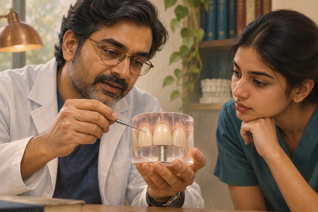
A successful anterior implant must support a crown whose outline, colour and mucosal margin fit the patient’s smile. Those goals depend on bone, mucosal phenotype, the location of the fixture and the shape of the restoration. The most consequential decisions often occur before surgery. A provisional crown can refine tissue contour, while an excessively facial implant axis or missing facial wall may require a different solution.
Separate the tissue variables
Record midfacial mucosal level, thickness, keratinized mucosa width, vestibular depth, papillae and the relationship to adjacent teeth. A band of keratinized tissue can be thin or thick. A thick phenotype does not guarantee a broad band of keratinized tissue or comfortable cleaning. Adjacent periodontal support and the contact area influence the appearance of the papilla. An isolated recession defect with otherwise healthy tissues presents a different problem from inflammation with progressive bone loss.
The peri-implant epithelial and connective tissue interface protects the bone beneath it. Collagen fibres next to the implant do not insert into cementum as they do at a natural tooth. Healthy probing depths vary with tissue dimensions and implant position. Document baseline depths and radiographs after rehabilitation so later changes can be interpreted in context. [5]
Design the contour in three dimensions
The intended crown defines a cervical outline, proximal contacts and a path for cleaning. Fixture depth and facial–palatal position determine how much vertical and horizontal room is available to make that transition. A shallow or facial platform can force an abrupt bulge; excessive depth can hide margins and make probing or cement removal difficult. Look at the implant platform relative to the planned crown and crest, and check it against the adjacent roots and facial bone. [3] [1]
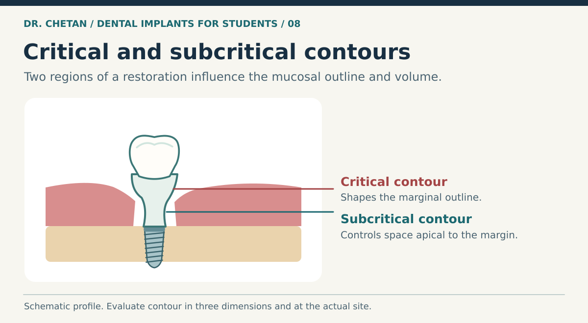
Critical contour describes the zone of the restoration near the mucosal margin that influences its level and outline. Subcritical contour lies apical to it in the transmucosal space. A provisional can be modified in these regions to guide the tissue. A more concave subcritical contour can provide space for tissue in selected situations. Excessive pressure or persistent blanching should prompt reassessment of the contour. Where the patient has a narrow vestibule, also check how the provisional affects hygiene access. [4] [3]
| Clinical finding | Main question | Planning implication |
|---|---|---|
| High smile line and asymmetric margins | How much tissue is visible, and what supports it? | Document expectations and compare the site with neighbouring teeth. |
| Thin facial tissue or a thin facial plate | Can the site preserve volume around the proposed fixture? | Consider staged care, tissue augmentation or alternatives when indicated. |
| Tenderness or difficulty cleaning at a site with limited keratinized mucosa | Is the tissue condition affecting comfort or plaque control? | Assess restoration contour, hygiene technique and whether augmentation is justified. |
| Overcontoured restoration | Is the problem due to position, contour or both? | Determine what can be changed restoratively before planning surgery. |
Select grafting for a defined endpoint
A connective tissue graft can add volume in selected thin or deficient sites. A free gingival graft can be useful where increasing keratinized mucosa or improving the hygiene environment is the principal aim. Substitutes can reduce donor-site morbidity and may be options in selected indications. Colour match, thickness, patient symptoms, cleaning access and stability over time are different outcomes. Evidence and recommendations depend on the defect, baseline conditions and aim of treatment. [2]
Transfer a successful provisional contour
The provisional offers a working record of the emergence profile after the tissues have adapted. At the definitive impression or scan, document the contour and its relation to the mucosal margin and adjacent teeth. The tissue can change shape soon after the provisional is removed, so the workflow should capture both the implant position and the contour that supported the tissue. The laboratory then needs enough information to reproduce or intentionally change the profile. At delivery, compare how the definitive restoration seats, supports the margin and accepts cleaning aids. A close colour match alone does not confirm that the contour was transferred. [3] [4]
A clinical example
At a healed anterior implant, the facial mucosa is thin and the provisional crown creates a sharp facial convexity. Before planning a graft, check fixture position and tissue health. If the fixture is appropriately positioned, changing the provisional contour may create space for tissue or clarify how much volume is missing. If the fixture is positioned far facially, contour adjustment and grafting each have limits. Discuss the esthetic aim, plausible outcome and maintenance with the patient. See planning and anatomy, immediate placement and peri-implant disease diagnosis.
References and further reading
- International Team for Implantology. Buccal bone wall thickness and peri-implant dimensional change
- International Team for Implantology. Medium- and long-term outcomes after soft tissue augmentation
- International Team for Implantology. Influence of restorative procedures on esthetic outcomes
- Su et al. Critical and subcritical contours of implant abutments and crowns
- Berglundh et al. World Workshop consensus report on peri-implant diseases and conditions

Leave a Reply