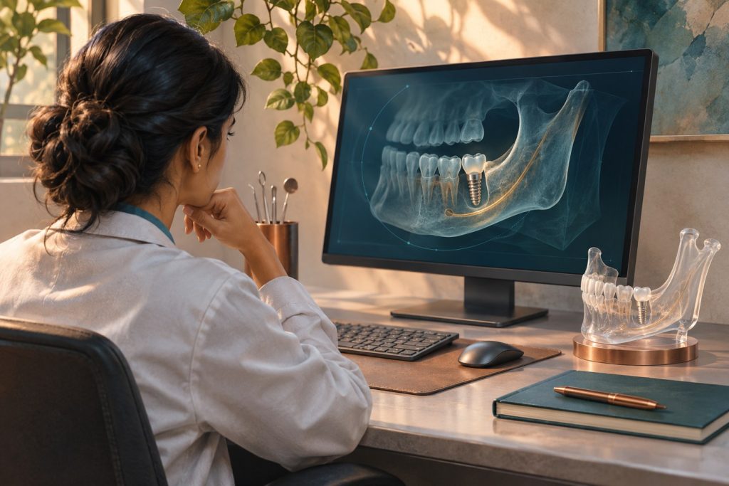
A ridge can accommodate a fixture while offering a poor position for the final crown. An implant placed too facially may require an overcontoured restoration or leave a thin facial plate. One placed too lingually can produce a ridge lap that the patient cannot clean. Prosthetically driven planning begins with the planned replacement and tests whether a safe, maintainable implant position can support it.
Plan the crown before testing the bone
Start with the tooth arrangement, incisal or occlusal plane, opposing dentition and proximal contacts. Identify where the screw channel would emerge if the restoration were screw-retained. Check interarch restorative space and the intended emergence profile. A diagnostic wax-up or digital design makes these decisions visible. For a full arch, assess lip support, tooth display, phonetics and the intended transition from prosthesis to tissue. Decisions made at this stage may change implant distribution or the entire restorative plan.
Use the clinical examination and existing radiographs to frame the imaging question. The 2026 ADA and AAOMR recommendations describe panoramic imaging for initial implant assessment and CBCT for presurgical planning and placement. Justify exposure for the clinical task, use the smallest field that answers it and arrange qualified interpretation of the captured volume. Clinical examination still supplies information about tissue mobility, the mucogingival junction and available opening. [1]
Read a volume as a surgical corridor
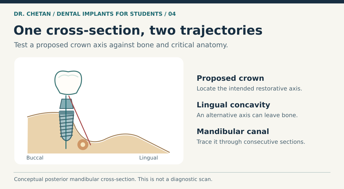
Inspect multiplanar slices, including cross-sections aligned to the proposed implant path. A panoramic reconstruction can locate a site, while cross-sectional views show width, concavities and the relationship of the drill path to surrounding structures. Trace relevant structures across consecutive slices. Avoid equating CBCT grey values with calibrated medical CT Hounsfield units or using a single intensity value as proof of bone quality. [6] [2]
| Region | Structures to follow | What changes when clearance is limited |
|---|---|---|
| Posterior mandible | Inferior alveolar canal, adjacent roots and lingual undercut | Reassess length, angulation and risk of lingual perforation or nerve injury. |
| Premolar and anterior mandible | Mental foramina and any anterior loop, incisive canal where relevant, lingual contours | Verify individual anatomy across slices and plan the usable corridor. |
| Anterior maxilla | Facial plate, adjacent roots, incisive canal and nasal floor | Assess three-dimensional emergence, facial bone and the true root-level space. |
| Posterior maxilla | Sinus floor and septa, residual crest, adjacent roots and relevant sinus findings | Reassess the implant plan, graft alternatives and need for additional evaluation. |
Where digital guides help and where errors accumulate
Merging surface scans with CBCT can bring the diagnostic tooth arrangement and bone into a single workspace. The quality of the registration matters. Metallic artefact, poor landmarks, a poorly fitting or rocking guide, sleeve tolerance and limited opening can all affect the transfer of a plan. Static guidance has measurable deviation at the entry, apex and axis. ITI guidance recommends allowing a safety margin from critical anatomy, with 2 mm discussed for static guided placement. That value is a planning buffer from the cited context. The clinician still considers the anatomy, system and uncertainty of each case. [3] [4]
Verify guide seating and inspect the proposed trajectory independently. A drill or implant may need more vertical access than a sleeve appears to require on screen. If the guide cannot be fully seated or verified, pause and reassess the method. Record the intended position, the anatomical constraints and the circumstances that would lead you to alter or defer the plan.
Read the position in three directions
Mesiodistal planning includes root clearance and the contact shape of the future crown. Buccolingual planning includes the facial plate, the canal or undercut where relevant and the position of the crown over the ridge. Apicocoronal planning considers the implant platform relative to the bone, available mucosal height and room to make the restorative transition. Evaluate all three together. Moving a fixture apically to create room for the emergence contour may complicate hygiene or access. Changing angulation to avoid a root may change the screw exit and the position of the apex. A useful treatment record shows these linked changes on the scan and restorative plan rather than recording only an implant length and diameter. [3]
A clinical example
A mandibular second premolar is missing. The proposed crown looks centred from the occlusal view. In the cross-section, the implant axis directs the apex towards a lingual concavity. Rotating the fixture to follow the ridge would place the access channel at the edge of a cusp. The team can compare alternative implant geometry, restorative design or a different treatment by looking at the crown, path and bone together. The objective is a position that remains safe to place and practical to restore and maintain. The patient assessment chapter frames the alternatives, and implant placement explains what must still be checked at surgery.
References and further reading
- Benavides et al. ADA and AAOMR recommendations for patient selection for dental radiography and CBCT, 2026
- International Team for Implantology. Cone beam computed tomography
- International Team for Implantology. Computer-guided implant surgery
- International Team for Implantology. Accuracy of static computer-aided implant surgery
- International Team for Implantology. The SAC Classification in Implant Dentistry, second edition
- Systematic review of CBCT grey values and Hounsfield units

Leave a Reply