An implant assessment starts with a restorative problem and the person who has it. A patient might want to chew on one side, avoid a removable appliance or improve a visible gap. Those goals can point to different treatments even when the radiographs look similar. Begin by defining the problem, assessing whether existing teeth can be maintained and comparing feasible alternatives. Treating a disease that threatens a tooth can change the decision to extract it.
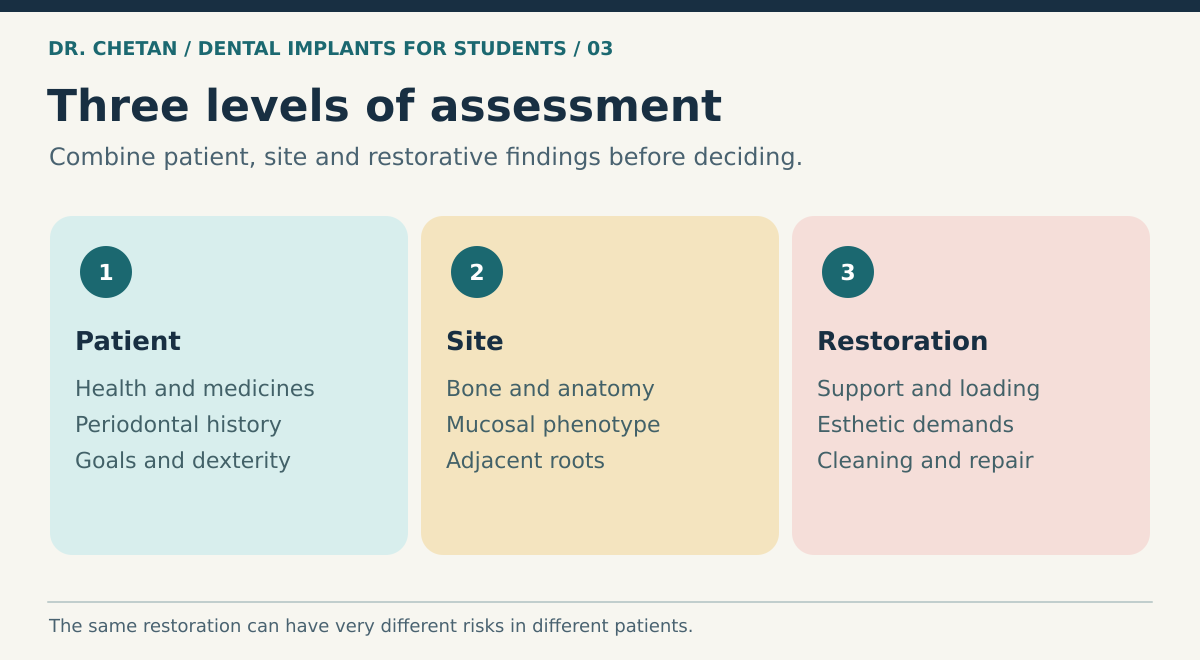
Assemble risk at three levels
| Level | What to document | How it changes the plan |
|---|---|---|
| Patient | Medical history, medications, tobacco exposure, glucose control, dexterity, attendance and priorities | May change healing expectations, preparation, referral and follow-up. |
| Site | Periodontal condition, bone envelope, soft tissue, adjacent roots, gap and anatomy | Determines position, need for augmentation and technical difficulty. |
| Restoration | Space, support distribution, force, esthetic demand, cleansability and repair access | Determines whether the final restoration can meet the agreed goals. |
Periodontal charting establishes the health of remaining teeth and the patient’s baseline capacity to control biofilm. Treat active periodontitis before elective implant treatment and verify a stable situation. A history of periodontitis remains relevant to maintenance even after successful treatment. Ask about smoking and discuss cessation support. Assess diabetes in context, including available measures of glycaemic control and complications. The presence of a diagnosis alone gives less information than its severity, treatment and stability. [2] [3]
When history changes the team or timing
Record the indication, route, duration and timing of antiresorptive and antiangiogenic therapy. Cancer-dose regimens and osteoporosis regimens have different risk profiles. The AAOMS position paper advises avoiding implant placement in oncology patients receiving parenteral antiresorptive or antiangiogenic therapy, with specialist management and individualized counseling for other settings. Previous radiotherapy involving the jaws warrants specialist review of the site, radiation history and risk of osteoradionecrosis. The dental team should coordinate medication decisions with the prescriber. [4]
Anticoagulation requires assessment of the intervention and bleeding risk and may call for consultation under the applicable protocol. An adult with controlled medical conditions may still have a highly complex implant site. In a young person, residual craniofacial growth can leave an implant crown infraoccluded relative to adjacent teeth. Estimating growth and explaining future changes can be more useful than applying a fixed age cutoff.
Assess difficulty before offering a procedure
The ITI Straightforward, Advanced and Complex framework separates surgical and restorative demands and uses modifiers to recognise when complexity rises. A broad healed posterior ridge and an uncomplicated single crown present different challenges from an immediate anterior implant in a thin phenotype with a high smile line. The latter demands coordination of extraction, hard and soft tissue management and provisionalization, plus a fallback if the plan cannot be achieved. Operator experience and access to a multidisciplinary team form part of the assessment. [1]
Discuss the whole treatment pathway. Outline alternatives, including retaining the tooth when feasible, a tooth-supported or resin-bonded bridge where suitable, a removable restoration or leaving the space in selected circumstances. Explain how provisional support, grafting, time, biological complications, mechanical repairs and future costs differ. The conversation should cover which decisions may need to change at surgery and how the patient will manage during healing. Record what the patient values, the agreed plan and the alternatives discussed.
Sequence disease control and restoration
Document the prognosis of the whole dentition. An isolated implant may seem straightforward until the opposing teeth, adjacent support and longer-term restorative needs are considered. Treat urgent disease, establish what can be preserved, then decide which missing sites need replacement. During planning, identify practical barriers to supportive care. A patient who can attend several surgical visits but cannot access follow-up near home needs a realistic handover and component record. Where treatment complexity rises, separate the decisions that can be made in general practice from those requiring input from a periodontist, prosthodontist, oral surgeon or medical clinician. [1] [2]
A clinical example
A patient with a missing upper lateral incisor requests a fixed implant tooth. The adjacent roots converge toward the gap and the patient has a high smile line. The crown appears to have adequate space on the cast. Root-level space and soft-tissue esthetics still require assessment. Orthodontic root repositioning, a resin-bonded bridge and an implant with a different timeline are all discussable. Showing the patient the proposed crown and the root relationship lets them weigh the consequences of each plan. The next step is a documented restoration-driven imaging assessment, informed by the esthetic tissue chapter.
References and further reading
- International Team for Implantology. The SAC Classification in Implant Dentistry, second edition
- Herrera et al. Prevention and treatment of peri-implant diseases. EFP S3 guideline
- AO and AAP consensus on peri-implant diseases and conditions
- American Association of Oral and Maxillofacial Surgeons. Position paper on medication-related osteonecrosis of the jaws, 2022 update
- U.S. Food and Drug Administration. Dental implant systems, components and materials

Leave a Reply