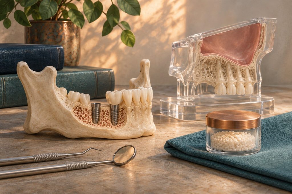
Bone augmentation should solve a defined restorative problem. A horizontal defect limits the width available for an implant and its surrounding bone. A vertical defect limits height and can create a long crown, difficult tissue transition or reduced support. Combined defects can change the achievable prosthetic design. Describe the defect against the intended implant position and crown before deciding whether to augment.
What makes a graft site capable of healing?
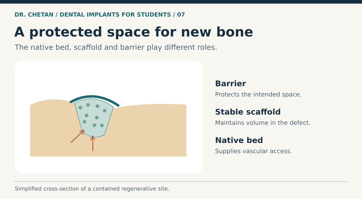
Guided bone regeneration aims to protect a space in which vascular and bone-forming tissues can develop. A barrier helps limit the early migration of soft tissue into the defect. The space needs support, blood supply and wound stability. The PASS framework describes primary wound closure, angiogenesis, space maintenance and stability. These principles explain why a membrane and graft material are useful only if the surrounding wound can support them. [2] [1]
Graft terminology describes biological roles. Osteogenesis is bone formation by living cells. Osteoinduction refers to signals that can drive precursor cells toward bone formation. Osteoconduction refers to a scaffold along which new bone can grow. Autogenous grafts can contribute living cells and biological signals, with donor-site morbidity and variable remodelling. Processed allografts, xenografts and synthetic substitutes differ in composition, turnover and volume stability. The material’s category alone gives limited information about the behaviour of a particular product.
Fit the procedure to the defect
| Defect and site | Options to compare | Main question |
|---|---|---|
| Contained gap or local facial deficiency | Particulate graft, membrane when indicated, or a different plan for the site | Will stable tissues and native bone support the desired result? |
| Horizontal ridge deficiency | Staged or simultaneous guided regeneration, block or other augmentation, selected narrower fixtures | Can the fixture be placed in the right position with adequate stability and coverage? |
| Vertical or combined deficiency | Staged or advanced reconstruction, a different prosthetic design or referral | Do the likely gain and maintenance justify the greater biological and technical demands? |
| Inadequate posterior maxillary height | Transcrestal or lateral sinus elevation, selected short implants or alternative rehabilitation | What volume is needed, and can the remaining bone provide stability? |
Simultaneous augmentation can reduce the number of surgical stages if the implant has adequate native-bone support and the defect can be protected. In a staged approach, the team reconstructs the ridge and reassesses after healing. Wound exposure, infection, donor-site symptoms, resorption and incomplete gain should be included in the treatment discussion. Persistent exposure can change the prognosis and management, especially when there is infection or graft instability. [1]
What changes in the posterior maxilla?
The Schneiderian membrane lines the sinus. Sinus floor elevation creates a protected space above the existing floor and below that membrane. A transcrestal approach accesses the floor through the crest, while a lateral window provides another access route and direct visibility of more of the membrane. The choice depends on residual height and width, the amount and distribution of gain required, sinus anatomy and the feasibility of stable placement. Examine septa and relevant sinus findings on imaging. Persistent sinus symptoms or clinically significant pathology may require additional evaluation. [3]
Membrane perforation is a recognised complication. Its extent, location and ability to be managed influence whether the plan can proceed. Short implants can be an alternative to sinus augmentation in some posterior maxillary sites. Compare the specific evidence, proposed crown geometry, force distribution, morbidity and follow-up. [4] [5]
Evaluate what the graft gained
Radiographic height and width are useful planning measures, but the outcome also includes wound healing, maintenance of volume, the possibility of placing the fixture in the intended position, the patient burden and the final prosthesis. A study reporting implant survival after successful reconstruction may give limited information about patients whose first augmentation failed or who chose another treatment. When explaining options, include the possibility of staged reassessment. A sinus elevation can increase available height while leaving inadequate crest width or a poor restorative axis. Recheck the entire corridor instead of assuming that improving one dimension resolves the other constraints. [1] [4]
A clinical example
A patient missing an upper first molar has adequate ridge width but limited bone below the sinus. The proposed crown needs a stable position and acceptable occlusion. The team compares a short implant with a sinus-elevation plan by estimating available native support, restorative space and surgical burden. The patient values fewer operations, but that preference has to be considered alongside the findings and evidence for the site. Review CBCT anatomy and primary stability before deciding whether the implant and augmentation can be planned together.
References and further reading
- International Team for Implantology. Bone augmentation in localized alveolar ridge defects
- Wang and Boyapati. PASS principles for bone regeneration
- Cambridge University Hospitals. Sinus lift procedures and clinical considerations
- Short implants versus standard implants with sinus elevation. Systematic review of randomized trials with at least five years of follow-up
- Jung et al. ITI consensus on implant length, design and medications

Leave a Reply