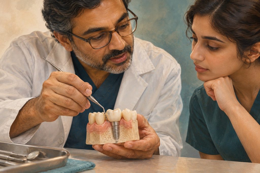
A deep probing measurement around an implant is a finding to interpret. Tissue thickness, position of the implant platform and the design of the restoration can influence the baseline depth. Diagnosis depends on inflammation and change over time, particularly the relationship between current probing and radiographic support. Baseline records taken after rehabilitation are part of the treatment.
What the case definitions actually require
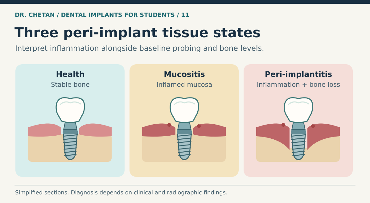
| Condition | Clinical findings | Bone findings |
|---|---|---|
| Peri-implant health | No erythema, swelling, bleeding on gentle probing or suppuration | Can be present around implants with reduced but stable support. |
| Peri-implant mucositis | Bleeding on gentle probing is the main sign. Swelling or suppuration may occur. | No bone loss beyond initial crestal remodelling. |
| Peri-implantitis | Bleeding and/or suppuration, increased probing depths compared with earlier examinations, and sometimes recession | Progressive bone loss beyond initial crestal remodelling. |
When previous clinical and radiographic records are absent, the 2017 World Workshop’s case definition for peri-implantitis uses a combination of bleeding and/or suppuration on gentle probing, probing depth of at least 6 mm, and bone levels 3 mm or more apical to the most coronal portion of the intraosseous part of the implant. These are criteria for a setting without baseline records. Where good baseline data exist, changes can support the diagnosis before a site reaches those values. [1] [4]
Examine the implant and its surroundings
Probe gently at accessible sites and record depths, bleeding or suppuration and recession. Compare readings with earlier records taken in a similar way. A standardized periapical radiograph can help compare mesial and distal crestal bone levels. Facial and lingual defects may be less visible in a two-dimensional image. Check whether the prosthesis obscures the examination. Assess plaque, restoration contour, residual cement where relevant, and whether the patient can clean the area.
If the prosthesis moves, localize the movement to the restoration, abutment or fixture. A truly mobile fixture suggests loss of integration. A loose restoration can alter probing access or irritate tissue. A single radiograph showing a low crest leaves unanswered whether the bone has changed since loading, how the platform was positioned and whether the implant is still healthy at the current level. See failure analysis for an approach to mechanical and biological findings.
Management follows the diagnosis and response
For mucositis, the EFP S3 guideline centres management on professional biofilm removal and effective home care, including correction of factors that obstruct access. Reassess the response. The diagnosis, patient’s ability to clean and the practical design of the restoration determine where to intervene. Routine systemic antibiotics are not a substitute for removing deposits and addressing the cause. [2]
For peri-implantitis, begin with a non-surgical phase that includes oral hygiene, risk-factor control, mechanical treatment and assessment of the prosthesis. Re-evaluate inflammation, depth and access. Sites with persisting disease may need specialist surgical assessment. Access, resective and reconstructive strategies address different anatomy and objectives. Defect configuration, maintainability and remaining support influence the choice. Long-term supportive care is needed even if the intervention reduces inflammation. [2] [3]
Reassess change rather than relying on one result
Use comparable probing and radiographic records to judge response. A site with less bleeding but persistent suppuration or continued bone loss needs further assessment. If oral hygiene has improved but a broad bridge still blocks access, a modification or removal of the prosthesis may be needed to examine and treat the site. Review the overall prognosis before choosing a reconstructive approach. A favourable defect alone cannot make an implant maintainable if the restorative position prevents cleaning. Referral is appropriate when diagnosis, access, surgical management or the decision to retain the fixture exceeds the team’s experience. [2] [3]
A clinical example
A molar implant had a probing depth of 4 mm at baseline and now measures 7 mm with bleeding and suppuration. A comparable radiograph shows new loss of supporting bone. This is a different clinical situation from a stable 6 mm site with no signs of inflammation and unchanged support. The longitudinal comparison, rather than the absolute depth alone, makes the diagnosis persuasive. Review mucosal anatomy and contours to understand baseline differences and maintenance to see how to collect comparable records.
References and further reading
- Berglundh et al. World Workshop consensus report on peri-implant diseases and conditions
- Herrera et al. Prevention and treatment of peri-implant diseases. EFP S3 guideline
- AO and AAP consensus on peri-implant diseases and conditions
- Renvert et al. Peri-implant health, mucositis and peri-implantitis case definitions

Leave a Reply