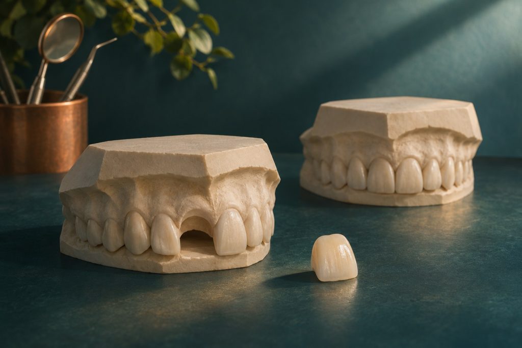
A fresh socket gives the surgeon an opportunity to combine visits and can help preserve the patient’s provisional tooth pathway. It also changes the geometry of the implant bed. The original root position often differs from the position that best supports the crown. A successful extraction leaves a site to assess again. The planned technique depends on the remaining socket walls, the native bone available for stability, and the restorative and tissue objectives.
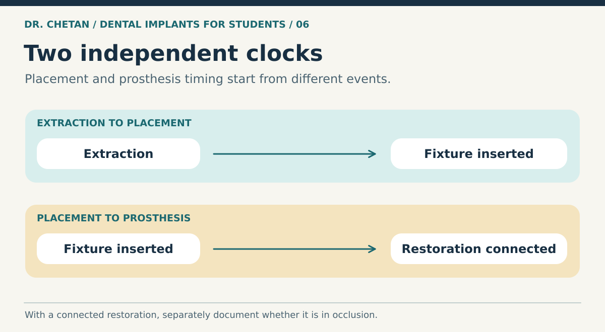
Use two independent clocks
| Decision | ITI terms | Practical meaning |
|---|---|---|
| Time from extraction to placement | Type 1 immediate, Type 2 early after soft tissue healing, Type 3 early after partial bone healing, Type 4 late after complete bone healing | Describes the condition of the site when the implant is placed. |
| Time from placement to restoration or loading | Immediate within one week, early from one week to two months, conventional after two months | Describes when a prosthesis is connected, with occlusal contact assessed separately. |
| Functional contact | Immediate restoration out of occlusion versus immediate loading with occlusal contact | Distinguishes early appearance or tissue shaping from intended functional loading. |
Type 2 commonly refers to placement after about four to eight weeks, and Type 3 after about twelve to sixteen weeks, in the cited ITI framework. These intervals describe the protocol used for classification and need interpretation alongside clinical healing. In the combined notation, Type 1A refers to immediate placement and immediate restoration or loading. Immediate placement with a conventional loading schedule is also possible. [1] [2]
Why a fresh socket changes the plan
Tooth extraction initiates remodelling, including reduction in facial ridge dimensions. An immediate fixture does not eliminate that response. A thin or damaged facial plate, thin mucosa, high smile line and pre-existing recession raise the esthetic stakes. The clinician evaluates the socket wall and defect, the facial gap, tissue contour and bone apical or palatal to the socket that could provide support in the required trajectory. A defect that cannot be managed predictably can change the choice to an early or staged plan. [3] [4]
In the anterior maxilla, the root axis may follow the facial plate more closely than the ideal restorative trajectory. Seeking stability simply by following that axis can place the fixture too facially. Position must support an emergence profile and accessible restoration. The ITI 2023 consensus also emphasises favourable selection and experienced teams for Type 1A. Selected sites with a chronic periapical lesion may be considered if conditions allow proper management and stability. An acute spreading infection or a site that cannot be adequately managed calls for a different approach. [2]
Provisionalization is a separate judgement
A provisional can guide soft tissue contour or close an anterior gap. Its shape and contact pattern must match the stability and protection planned for the wound. Occlusal contacts in maximum intercuspation and excursions, parafunction and the direction of expected force all matter. A restoration held out of contact may still receive incidental forces from food or the tongue. A removable or tooth-supported provisional can preserve appearance when the implant itself needs protection. Its fitting surface must also avoid pressure on the surgical site. [5]
When comparing survival rates, check whether studies report the extraction and loading timing clearly and how they dealt with patients who failed eligibility at surgery. Outcomes in a selected anterior cohort cannot be applied to every molar socket or every esthetic risk profile.
Evaluate the outcome beyond fixture survival
Immediate placement studies can report excellent survival while still finding midfacial recession, loss of tissue contour, or a need for further grafting or restorative modification. Ask which outcomes were measured and when. A short-term photograph cannot establish stable papillae or a maintainable restoration. A study that excluded sockets with a damaged facial wall answers a different question from a study of compromised sites. In an anterior case, document the initial mucosal margin, the appearance of adjacent teeth and the patient’s esthetic expectations so you can compare the result after maturation. [2] [4]
A clinical example
A fractured maxillary central incisor is planned for immediate replacement. After extraction, a substantial facial wall defect is visible and the proposed trajectory has little apical engagement. The patient expected a fixed temporary tooth. The team can change to a tooth-supported provisional and stage implant treatment, preserving the appearance plan while changing the surgical timing. That option should be discussed before extraction. See prosthetic planning, augmentation and esthetic tissue management.
References and further reading
- International Team for Implantology. Implant placement and loading protocols
- Morton et al. ITI Group 5 consensus report on implant placement and loading protocols, 2023
- International Team for Implantology. Implants in postextraction sites
- International Team for Implantology. Buccal bone wall thickness and peri-implant dimensional change
- International Team for Implantology. Loading protocols for single implants

Leave a Reply