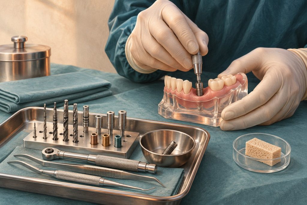
The surgical appointment tests whether the plan made on the model and scan can be achieved in living tissue. The desired result is an implant in a safe restorative position with a viable wound, adequate initial support and an appropriate healing plan. Surgical teaching should therefore connect each action to an objective and a reason to stop or change course.
Preserve the site while transferring the plan
Before treatment, verify the site, the planned restoration, imaging findings, components, consent and the backup provisional. The approach determines how much of the anatomy can be seen. A flap can expose the crest or allow augmentation, at the cost of additional tissue manipulation. A flapless approach can preserve soft tissue in carefully selected circumstances but offers limited direct view of the ridge. Whether a guide is used, the surgeon must be able to verify seating, access and orientation. [1]
Osteotomy preparation shapes a corridor for the fixture. Its direction must serve the restoration and avoid structures identified in the plan. Cutting efficiency, irrigation, drill wear, surgical access and pressure affect heat generation and bone injury. Protocols vary with implant geometry, instrumentation and site conditions. Manufacturers supply system-specific guidance. Underpreparing a site can increase insertion resistance in some situations, but excessive compression of dense cortical bone can compromise the tissue expected to heal. The aim is sufficient engagement in the right position with viable bone at the interface.
Interpret the stability measurements together
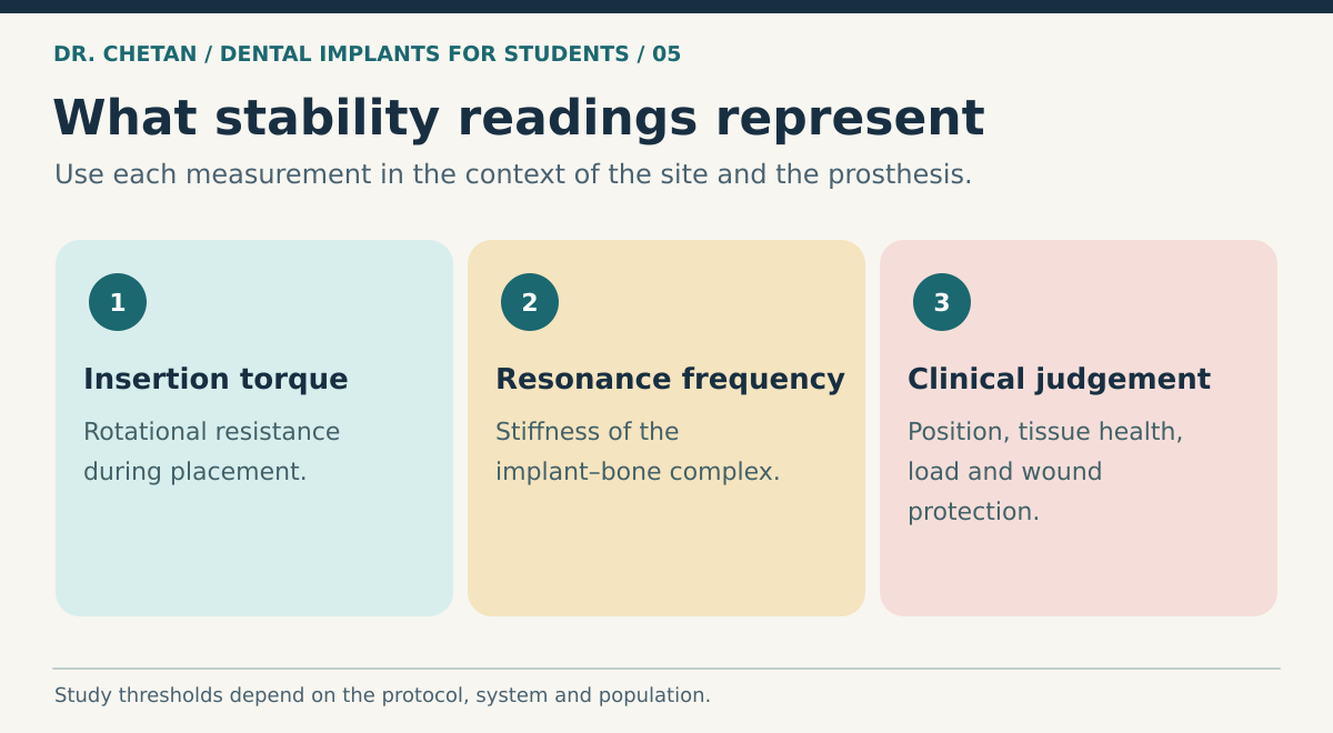
Insertion torque reflects rotational resistance during placement. Resonance-frequency analysis evaluates the stiffness of the implant–bone system and reports an implant stability quotient. Transducer orientation, bone support, exposed implant height and measurement conditions can influence the reading. Values from unlike sites or differently oriented measurements are difficult to compare directly. Document the device, orientation and clinical setting if serial readings are being used. [2]
| Finding at placement | Possible implication | Decision that follows |
|---|---|---|
| Adequate engagement in the planned position | Mechanical support is present at insertion | Reassess the tissue, restoration and loading plan together. |
| High rotational resistance in dense bone | Strong engagement or excessive compression | Review the preparation and system guidance, and evaluate seating. |
| Low engagement or an unexpected defect | Inadequate support for the original plan | Consider protection, staged treatment or deferral based on the findings. |
| Stable fixture with a major adjacent graft | Fixture and regenerated tissues have different healing needs | Plan wound protection and loading around both. |
Torque or ISQ values reported in selected studies belong to those protocols. The evidence does not support treating a single number as a universal permission to load. Review planned occlusal contact, parafunction, grafting, the consequences of micromotion and whether an alternative provisional can be used. The healing biology chapter explains why mechanical resistance on day one differs from later integration. [3] [4]
Wound management and the early review
A transmucosal healing component can shape an accessible opening during integration. A submerged cover screw may be selected where tissue coverage or graft protection is important and requires later exposure. A healing abutment and an implant-supported provisional are distinct components with different functions. Whether a temporary restoration is fixed or removable, check for unintended contact with the wound or implant.
At review, assess the wound, tissue closure, symptoms and provisional. Record baseline sensory findings if the anatomy or symptoms warrant it. Progressive swelling, spreading infection, persistent uncontrolled bleeding or new altered sensation require assessment appropriate to the urgency. Do not assume that a quiet wound proves integration. Coordinate restoration timing with the clinical assessment of healing and site stability. [5]
Document the difference between plan and finding
Record fixture identity, dimensions, site, position, primary stability findings, graft and membrane if used, and whether the implant was left submerged or transmucosal. Describe material deviations from the plan and explain how they change the next visit. If a planned implant could not be placed, document the anatomical or biological finding and the temporary arrangement offered. A restorative colleague who receives this record can distinguish a stable fixture awaiting routine review from one whose loading decision remains unresolved. In clinical teaching, this handover is part of the procedure because it determines how the next clinician interprets the site.
A clinical example
A planned posterior implant achieves adequate engagement, but an unexpected facial dehiscence is found at placement. An immediate provisional would interfere with the protection of the grafted area. The initial fixation answers one question. Tissue coverage and the ability to keep the provisional away from the site answer others. A protected healing plan may better fit the changed situation. See bone augmentation and placement and loading after extraction.
References and further reading
- International Team for Implantology. Computer-guided implant surgery
- Resonance frequency analysis and clinical outcomes. Systematic review and meta-analysis
- International Team for Implantology. Loading protocols for single implants
- International Team for Implantology. Implant placement and loading protocols
- Herrera et al. Prevention and treatment of peri-implant diseases. EFP S3 guideline
Leave a Reply