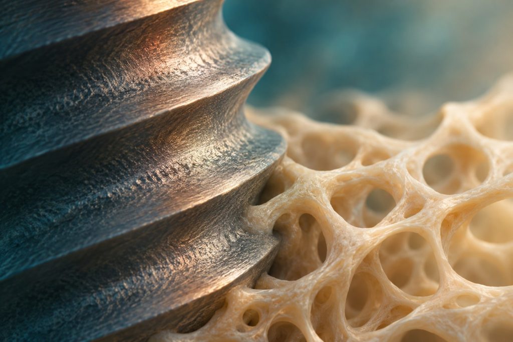
Implant placement creates a wound in living bone. The prepared site provides immediate mechanical support, while damaged bone, blood and the implant surface form a new healing interface. The subsequent sequence explains why a fixture that feels stable on the day of placement can still be vulnerable in the following weeks, and why a clinically quiet implant is assessed over time.
From a surgical wound to a load-bearing interface
Osseointegration describes the direct structural and functional relationship between living bone and a load-bearing implant surface. The interface is heterogeneous. In histological sections, some regions show direct bone contact, while other regions contain marrow or gaps. Bone-to-implant contact is a histological measurement over the assessed surface. A periapical image or a resonance-frequency reading cannot report that percentage in a living patient. [2] [1]
The early sequence begins with bleeding and the adsorption of proteins to the surface. Platelet activation and a fibrin network form a provisional matrix. Inflammatory cells clear debris and release signals that help recruit repair cells. Capillaries and osteogenic cells enter the healing space. New bone appears as woven bone before remodelling produces a more organised architecture. Bone turnover continues after loading.
Distance osteogenesis describes bone formation progressing from the cut bone surface toward the fixture. Contact osteogenesis describes new bone formation along the implant surface after cells migrate to and colonise it. Both can contribute in the same site. Their relative expression varies with the local gap, vascularity, surface and healing conditions. Early bone formation at an implant surface is therefore a biological response to a wound and material interface. [1]
Why two kinds of stability matter
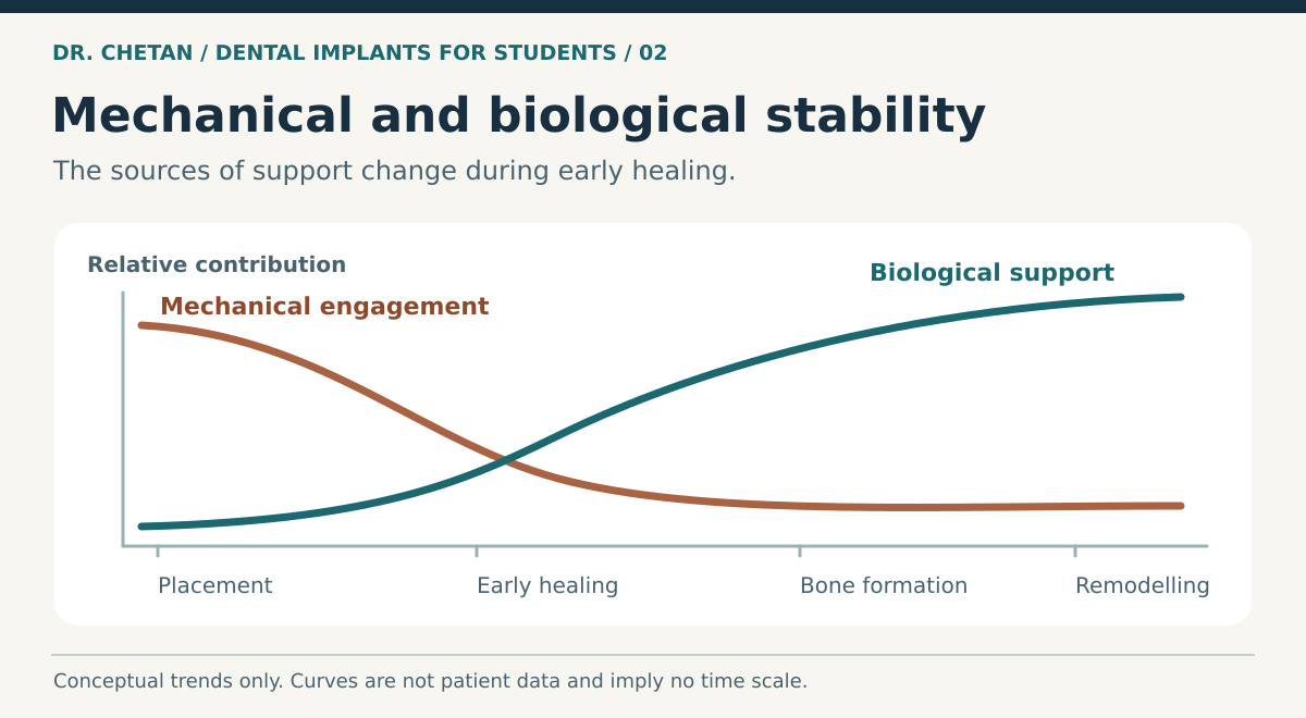
Primary stability is the mechanical restraint present at insertion. Cortical engagement, bone geometry, trabecular density, fixture macrogeometry and site preparation influence it. Secondary stability comes from new bone formation, maturation and remodelling at the interface. Existing bone involved in primary engagement can remodel while secondary stability is developing. Some sites show a temporary reduction in a measured stability signal during that transition. The timing and size of the change vary. [3] [4]
| Observation | What it represents | What it cannot establish |
|---|---|---|
| Insertion torque | Rotational resistance during insertion | Whether new bone has formed or whether immediate loading is appropriate on its own. |
| Implant stability quotient | A resonance-frequency-derived measure of stiffness of the implant–bone complex | Histological bone contact or guaranteed survival. |
| Clinical and radiographic review | Evidence about symptoms, mobility, bone levels and change over time | A direct measurement of the cellular events at the interface. |
A roughened surface can influence clot adhesion, cell behaviour and the rate of early bone response. Thread shape and taper chiefly affect the mechanical interaction during placement. Marketing descriptions often combine them. When reading a study, identify which property changed and whether the outcome was a histological measurement, a stability reading or clinical survival.
How healing fails
Sustained excessive relative motion at the interface can favour fibrous repair rather than stable bone attachment. The relationship depends on load magnitude and direction, its repetition, geometry and the quality of the healing site. A single micromotion number from an experimental model cannot be used as a universal clinical threshold. Overheating, inadequate blood supply, excessive compression, contamination and patient factors can also interfere with healing. Several causes can coexist in the same failure.
A natural tooth is suspended by a periodontal ligament that contributes to load distribution and sensory feedback. An implant transfers load more directly through its bone interface. That difference helps explain why restorative position, occlusion and force management matter at insertion and beyond. The tissue response is best understood alongside prosthetically driven planning and the restoration chapter.
Timing the evidence
Early healing research uses animal histology, experimental interface models and clinical proxies. Their outcomes answer different questions. A higher early percentage of contact in a histological specimen can suggest a faster bone response under the conditions studied. It does not by itself show that a new design reduces long-term loss, shortens treatment in all sites or changes the threshold for loading. Likewise, an unchanged radiographic crest does not reveal how much of the surface is covered by new bone. When comparing studies, identify the species or patient group, surface, sampling time and endpoint before applying a result to clinical care. [1] [4]
A clinical example
Two implants have the same insertion torque. One is in a healed site with a favourable axis and healthy soft tissues. The other relies on a small area of native bone beside a grafted defect. The number is similar because it measures rotational resistance at insertion, but the healing environment, force directions and consequences of early loading differ. The team may load them at different times. Asking which tissue supports the implant today, which tissue must heal, and how the planned provisional would transmit force gives a more informative assessment than comparing the readings alone. See placement and surgical healing for how those findings influence the operative plan.
References and further reading
- Davies JE. Understanding peri-implant endosseous healing
- Albrektsson et al. Requirements for direct bone anchorage of titanium implants
- Raghavendra, Wood and Taylor. Early wound healing around endosseous implants
- Resonance frequency analysis and clinical outcomes. Systematic review and meta-analysis

Leave a Reply