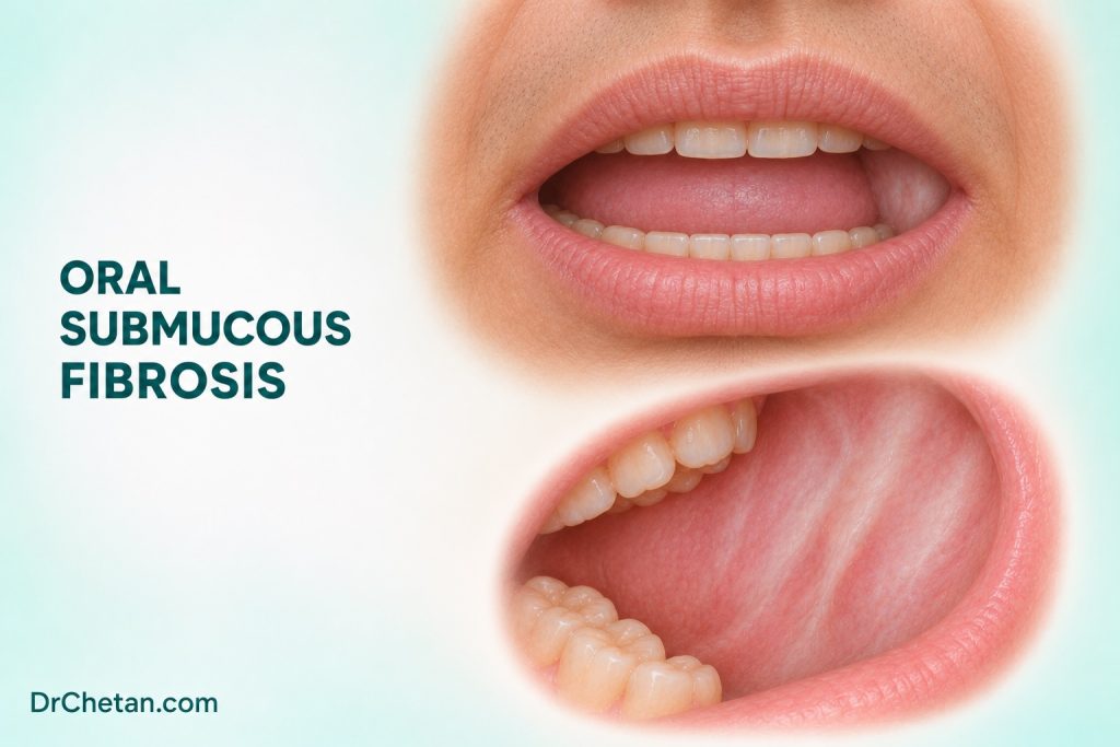
A patient reports burning with spicy food and says it has become harder to open their mouth. Examination shows pale, less pliable buccal mucosa and palpable bands. The patient uses flavoured supari but has never smoked or added tobacco to it. This combination raises suspicion for oral submucous fibrosis (OSF), an oral potentially malignant disorder strongly associated with areca nut. The clinician needs to document the functional change, examine for a focal lesion and arrange appropriate assessment, especially when restricted opening limits the view.
Connect the symptoms with changes in the mucosa
OSF is a chronic fibrotic disorder of the oral mucosa. Burning, sensitivity to spicy food and a sense of stiffness can precede marked restriction in opening. As disease progresses, the mucosa may appear blanched, feel less pliable and develop palpable fibrous bands. Buccal mucosa, labial mucosa and soft palate may be involved, with variable effects on tongue mobility and other structures. Symptoms, distribution and severity vary. Record what the patient can do and which parts of the mouth you can examine.
Ask about areca nut by product name. Supari, pan masala and betel quid can contain areca without tobacco, while gutka and some quids contain both. Ask separately about frequency, years of use, typical placement and whether tobacco is added. Record any past cessation or change of product. Areca exposure supports the clinical assessment and provides a specific prevention target. Tobacco, when present, adds another exposure to address.
Examine the whole mouth and record a functional baseline
| Finding | What to record | Why it matters |
|---|---|---|
| Burning or intolerance to certain foods | Onset, location, triggers and progression | Helps distinguish symptom burden from visible severity and guides follow-up questions. |
| Blanching and reduced mucosal pliability | Sites affected, symmetry and associated surface changes | Shows the distribution and helps identify an abnormal focus within diffuse change. |
| Fibrous bands | Palpable sites, side and relation to the labial or buccal mucosa or palate | Supports the clinical impression in the appropriate context. |
| Reduced opening | Measured interincisal distance, landmarks used, discomfort and examination limits | Provides a reproducible baseline and identifies sites that could not be adequately assessed. |
| New focal red or white change, ulcer, mass or firmness | Exact site, size, progression and any associated neck findings | Can require prompt separate diagnostic assessment. |
Measure maximum interincisal opening in millimetres using identifiable upper and lower incisal edges when present. If dentition is missing or altered, record the landmarks and method used so the measurement can be compared later. Note whether pain, muscle guarding or another factor limited the effort. Several OSF staging systems use different thresholds and clinical features. A measured value and a clear description of function are useful even when the receiving team uses a different system.
Restricted opening has a wider differential. Temporomandibular joint disorders, muscle spasm, infection, trauma and other causes can restrict opening. Consider the time course, symptoms and examination alongside the mucosal findings and areca history. Abrupt restriction with significant pain or infection findings requires assessment for the acute process. A patient can have a fibrotic disorder and an independent cause of new functional change.
Record limitations during examination
If the patient cannot open enough to see the posterior buccal mucosa or retromolar region, record that before describing the rest of the examination. Ask about symptoms in the areas that remain out of view and assess any neck findings within your role. A note such as “posterior right buccal mucosa incompletely visualised because of restricted opening” tells the receiving clinician what remains to be assessed.
Understand the histopathological features

Early samples can show inflammation and changes in the subepithelial connective tissue. As fibrosis develops, collagen can become more dense and hyalinised beneath the epithelium. More advanced examples can show reduced vascularity, less cellular fibrotic tissue and involvement of deeper tissues. The overlying epithelium may be atrophic, and its appearance can vary across sites. These changes depend on the area sampled and the stage of disease. A single histological feature does not replace the clinical description of mucosal stiffness, bands and measured opening.
The mechanisms involve abnormal matrix deposition and turnover. Areca-derived compounds can affect fibroblast activity and collagen regulation. Inflammatory and profibrotic signalling contribute to a tissue environment in which fibrosis can persist. This helps link the microscopy to reduced pliability and restricted movement. Mechanistic research does not give a chairside test that can predict an individual patient’s progression or cancer risk.
Check for epithelial dysplasia and invasion separately from the degree of fibrosis. The pathologist may report dysplasia in an overlying epithelium or invasive carcinoma in a focal lesion. A sample confirming fibrosis from one site cannot establish the tissue diagnosis of a different ulcer or firm focus. When focal findings are suspicious, the receiving clinician decides how to assess the specific area and how to obtain an adequate sample given the limits of access.
Decide what needs biopsy or specialist referral
The clinical pattern and exposure history can strongly support OSF, and specialist assessment or biopsy can be useful when the presentation is uncertain. The reason for biopsy should be clear. A sample intended to evaluate background fibrosis answers a different question from a sample targeting a persistent ulcer, new red patch, firm mass or other suspicious focus. Record the exact site, whether it can be fully seen and which features prompted concern.
OSF is associated with an increased risk of oral cancer. Systematic reviews have found malignant transformation across studied cohorts, with variation in case definitions, geography, follow-up and selection of patients. A pooled estimate describes a study population. It does not establish an annual or individual risk. A new focal change warrants assessment based on its findings rather than on a quoted percentage. Continued examination matters even after a patient stops areca use.
Plan cessation, examination and ongoing review together
Explain that tobacco-free areca preparations can still cause harm. Ask which situations make the habit difficult to stop and agree a next step or offer cessation support appropriate to the product. Where tobacco is also used, address nicotine dependence separately. For the OSF itself, document burning, opening and daily function so the specialist has a baseline. Treatment decisions depend on severity, functional goals and available evidence. A Cochrane review found uncertainty for many interventions, and symptomatic or functional improvement and cancer surveillance are separate goals.
Set a review plan that includes the accessible mucosa and changes in opening, and state how the patient can return earlier if a new ulcer, red area, mass or neck lump develops. When examination is incomplete, name the surfaces that could not be seen and record how further assessment will be arranged. The responsibility for follow-through should be clear to the patient and referring clinician.
Describe what changes between visits
Compare the opening measurement using the same landmarks and method where possible. Ask about activities such as eating, speaking and maintaining oral hygiene, since two people with a similar measured opening can report different functional difficulties. Document the site and extent of bands and whether the areas previously out of view can now be assessed. A change in function can guide specialist review and rehabilitation, while each mucosal finding still needs its own clinical assessment.
When discussing the pathology report, identify whether it came from background mucosa or a focal lesion. The patient may hear “fibrosis” as a complete explanation for all their symptoms. Explain what that specimen can establish and which findings still need follow-up. If a specialist has planned a separate assessment of a focal change, give a clear appointment or contact route and record who will check that it occurred.
Two examples for case discussion
Burning and early functional change. A patient who uses areca daily reports burning when eating and notices that opening feels tighter. Examine the distribution of blanching and stiffness, measure opening and record any bands or focal mucosal changes. Discuss cessation and arrange assessment appropriate to the findings. The baseline lets the next clinician see whether the function or distribution has changed.
Restricted opening with an incompletely seen ulcer. A patient with known OSF reports a new unilateral sore and a change in discomfort. Only part of a focal ulcer is visible. Record what can be seen, the extent of the limitation and any palpable firmness or neck findings. Arrange prompt specialist assessment when the presentation is suspicious. A previous diagnosis of fibrosis does not establish the cause of the new lesion.

Leave a Reply