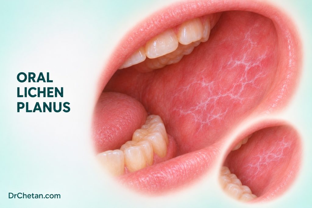
A patient has fine white lines on both cheeks and no symptoms. Another reports burning when eating and has a red erosion with white lines at its edge. Both appearances can occur in oral lichen planus (OLP), but the second visit also raises a practical question about what explains the focal erosion. Dental students should document the distribution, identify discordant findings, and use clinical and pathological information together.
Recognise the typical patterns
OLP is a chronic immune-mediated disorder that can involve the oral mucosa, sometimes with skin or other mucosal involvement. The oral findings can change over time. Fine reticular white striae are often bilateral and roughly symmetrical, especially on the buccal mucosa. Atrophic and erosive disease can cause burning or pain. More than one appearance can be present in the same mouth, and symptomatic red change can sit beside relatively quiet reticular areas.
| What to look for | What changes the assessment | |
|---|---|---|
| Reticular | Fine intersecting white striae, often bilateral | Distribution, symmetry and whether a distinct plaque or firm focus sits within the background pattern. |
| Atrophic or erythematous | Red areas, sometimes around white striae | Focal progression, pain, bleeding and alternative explanations for redness. |
| Erosive | Superficial ulcerated or eroded areas, often painful | Persistent focal ulceration, induration or a lesion that no longer fits the rest of the mouth. |
| Plaque-like | Broader white areas, sometimes with striae elsewhere | A persistent isolated white plaque needs its own differential, including leukoplakia. |
| Desquamative gingivitis | Red, tender or peeling gingiva | This is a descriptive finding. Consider immune blistering disorders and other causes as well as OLP. |
Document the sites on both sides of the mouth, including the gingiva, tongue and buccal mucosa where relevant. Ask about onset, fluctuating symptoms, medicines and local contacts. If there are skin or other mucosal symptoms, record them and coordinate assessment within the appropriate specialty. A sharply localised lesion next to a restoration or a new focal change within bilateral disease can require a different explanation.
Use clinicopathological correlation
The American Academy of Oral and Maxillofacial Pathology position paper describes a combination of clinical and histopathological criteria. The clinical picture can include a multifocal, symmetrical distribution with characteristic appearances. A lesion limited to a local contact site or whose onset follows a plausible exposure needs additional correlation. The histology commonly includes interface inflammation with a predominantly lymphocytic infiltrate and basal cell injury. Similar changes can occur in other lichenoid disorders. Give the pathologist the distribution, clinical differential and the reason for sampling the chosen site.
A biopsy can help resolve an uncertain or atypical diagnosis, evaluate a persistent red or ulcerated focus, or assess possible epithelial dysplasia or invasion. A sample from a typical striated region and a sample from a new firm ulcer answer different questions. In patients with desquamative gingivitis, blistering or unexplained erosions, a specialist may arrange a separately handled specimen for direct immunofluorescence to investigate an immune blistering disorder. The diagnostic team selects the site and specimen handling for the tests it needs.
Diagnostic frameworks differ in how they handle dysplasia and lichenoid inflammation. The AAOMP framework uses the absence of epithelial dysplasia as part of its histopathological criteria for a classic OLP diagnosis. Other research addresses lichenoid presentations with dysplasia separately. When dysplasia is reported, record its grade, site and the specialist interpretation. A familiar clinical label should not obscure the management implications of a dysplastic or invasive focus.
Give the pathology team enough clinical context

A useful request can describe bilateral reticular striae across the buccal mucosa and specify that the sample came from a new red area on one side. Add the duration, symptoms and any suspected local or drug exposure. If a specimen is taken to investigate a possible immune blistering condition, communicate that question before sampling so the appropriate site and handling can be planned. A pathology result gains meaning from these clinical details.
When the report comes back, ask whether it explains the typical background pattern and the focal area of concern. Those can be separate questions. If the report describes an inflammatory pattern but the lesion remains firm or progressive, revisit the sample site and discuss further assessment. Record the action as well as the histological term.
Explain malignant potential with the right context
International consensus includes OLP among oral potentially malignant disorders. Systematic reviews report a low but measurable pooled proportion of malignant transformation, with estimates that change when case definitions, dysplasia handling and follow-up differ. A 2024 review pooled a transformation proportion of about 1.4% for studies classified as OLP. This is a study-level estimate across varying cohorts and observation periods. It does not give an annual risk or a patient-specific prediction. A separate 2026 review restricted to longitudinal studies of OLP without dysplasia at diagnosis reported a pooled estimate of 1.56%, with a 95% confidence interval of 1.14% to 2.12% and substantial heterogeneity. The reviews used different eligibility criteria, so their estimates are best read alongside those definitions.
Persistent red or erosive changes and new clinical discordance are reasons to reassess the individual finding. Site and exposures may contribute to risk in observational studies, but their absence does not resolve a concerning lesion. When discussing risk, explain why a new lump, persistent focal ulcer, firm area or unexplained growth merits assessment even in someone whose usual symptoms fluctuate.
Separate symptom relief from surveillance
Some people with asymptomatic reticular changes need explanation and follow-up, with treatment tailored to symptoms and findings. For symptomatic OLP, topical corticosteroids are commonly used under an appropriate clinician’s direction. A Cochrane review found low-certainty evidence of pain benefit over placebo for topical corticosteroids in the studies assessed. Management should include a plan to review response, consider adverse effects and reassess a lesion whose appearance changes. If symptoms persist despite treatment, review the diagnosis and consider other causes, including superimposed infection where appropriate.
Decide the follow-up interval with the treating clinician according to symptoms, clinical findings and pathology. Record the parts of the mouth that were examined, what has changed and who will review the patient. Give a contact route for earlier assessment when a focal lesion changes. Treatment of diffuse soreness can be useful while a separate suspicious area is investigated. Symptom improvement alone does not establish the diagnosis of a persistent focal lesion.
At the review, ask whether treatment changed pain, eating or tolerance of oral hygiene. Then examine the mucosa and compare the same sites with prior records. A patient may report less burning while an isolated ulcer remains. Record both outcomes and reassess the ulcer according to its current clinical features. If symptoms involve the skin, eyes or another mucosal site, help coordinate assessment with the appropriate service.
Two chairside examples
Bilateral striae without pain. An adult has fine symmetrical white lines on both cheeks, no ulceration and no focal firmness. Record the distribution, ask about symptoms and exposures, and discuss the assessment and follow-up plan with the supervisor. If the appearance or course later becomes atypical, use the baseline to identify which area changed.
Erosive disease with a new focal ulcer. A patient with previously documented bilateral reticular change develops a persistent firm ulcer on the lateral tongue. Describe the new finding separately, compare with earlier photographs or reports and arrange prompt specialist assessment or biopsy within competence. The background pattern and the new ulcer can require different clinical decisions at the same visit.

Leave a Reply