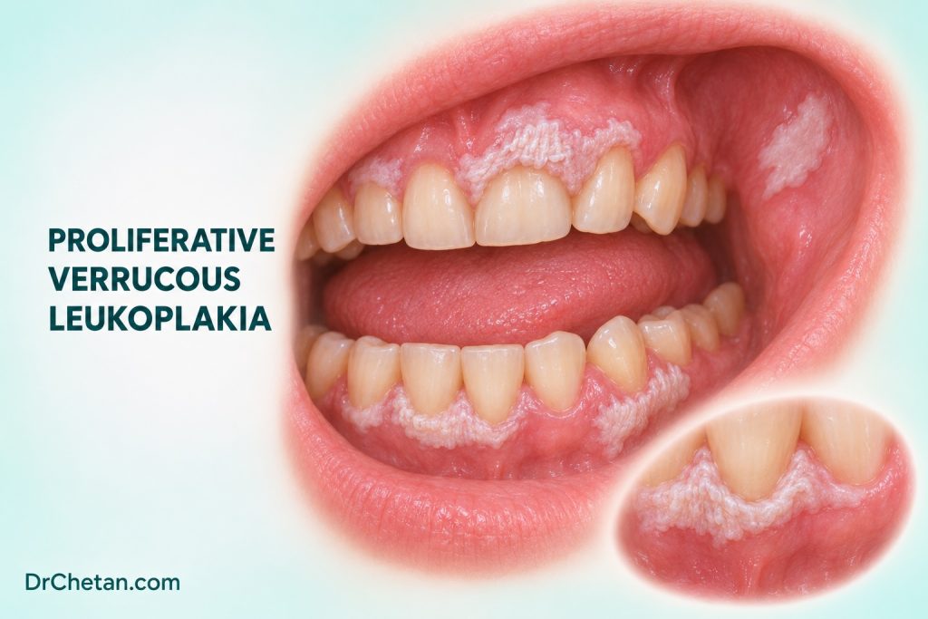
A patient has a small white gingival plaque. The first biopsy shows keratosis without dysplasia. On later visits, the original plaque broadens and a second plaque appears at a different site. One region becomes nodular. These changes are more informative together than any single photograph or pathology result. Proliferative verrucous leukoplakia, or PVL, is recognised through the evolving pattern across the mouth and its correlation with serial pathology.
Look for change across sites and visits
PVL is a potentially malignant disorder with a progressive course, often involving several oral sites. The early surface can be flat and white. It may later become thicker, corrugated, nodular or verrucous, and plaques can recur after treatment. Gingiva, alveolar mucosa and buccal mucosa are common sites in published cohorts. The sequence and speed of change vary. Some patients first present with limited disease, so an isolated early plaque may only become recognisable as part of a wider pattern over time.
Published series often include older adults and many women. PVL also occurs in other groups and in people without conventional tobacco exposure. Demographic features can support clinical awareness but cannot establish the diagnosis or exclude it. The history of progression, involvement of several sites and correlation between clinical and pathological findings carry more weight in an individual case.
| Finding | Useful interpretation | Next information to obtain |
|---|---|---|
| Single warty plaque | A surface description with several possible causes | Document the site, review local factors and obtain appropriate assessment if suspicious. |
| Similar plaques at several sites | A distribution that may fit progressive or reactive disorders | Map each site and compare with past records, examinations and samples. |
| Thickening or recurrence after treatment | A change that merits clinical comparison | Distinguish the treated site from a new site and review the prior histology. |
| New firm, red, ulcerated or enlarging focus | A concerning change that can coexist with less suspicious plaques | Arrange prompt assessment and identify which focus prompted it. |
Distinguish PVL from common mimics
A frictional keratosis may have a convincing contact source and a distribution that fits it. Lichenoid disease can produce bilateral white changes and erosions. Reactive or benign epithelial proliferations can also look warty. Conventional leukoplakia can have a verrucous area and may warrant its own risk assessment. A single verrucous plaque does not by itself demonstrate the progressive course of PVL. If the lesion has changed, reassess the current findings and review whether the earlier diagnosis still accounts for the overall pattern.
Verrucous carcinoma and conventional invasive SCC are especially important to consider when a focus becomes bulky, firm, ulcerated or progressively enlarges. A superficially sampled lesion can leave the deeper interface unevaluated. The clinical picture may justify further assessment even when the first sample was reported as keratotic. Communicate the degree of suspicion and the reason the previous sample may not answer the question.
Build a map another clinician can use

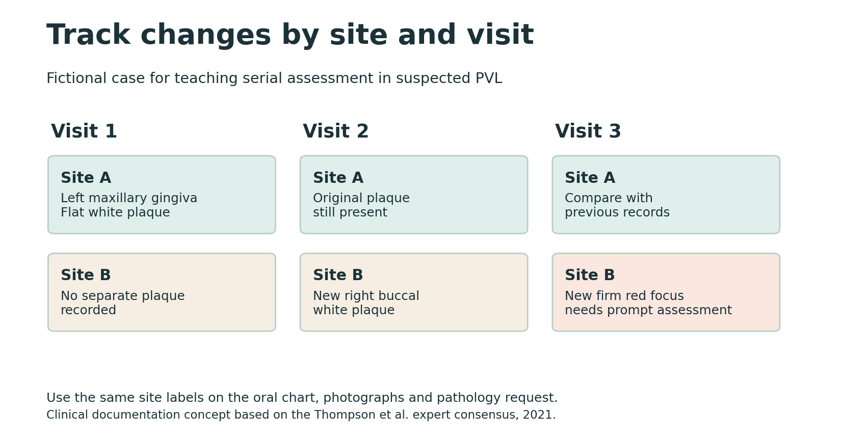
Assign each lesion a stable label, such as A for left maxillary attached gingiva and B for right buccal mucosa. Write the side, subsite, measurements and relation to landmarks. Record whether the plaque is flat, thickened, warty, mixed red-white, ulcerated or firm. If photographs are obtained with consent, use a consistent angle and scale where possible. Keep the earlier descriptions so that a new focus can be compared with an actual baseline.
The chart and pathology request should use the same lesion identifiers. A sample from “B, anterior edge of right buccal plaque” can later be distinguished from one from a thickened posterior focus in the same plaque. When a new lesion appears, add a new label rather than reusing a previous one. This lets the team distinguish extension of an existing lesion, change after treatment and a new focus.
Interpret the biopsy in the context of its site and time
The expert pathology consensus proposes standardised reporting language for lesions in the clinical context of PVL. Its categories cover corrugated keratotic lesions, bulky hyperkeratotic epithelial proliferations and findings suspicious for or diagnostic of SCC. The terms describe sampled tissue and support communication across serial biopsies. They are not required clinical stages that every patient must pass through in order. A specimen’s depth and the presence of a representative epithelial-stromal interface can materially affect interpretation.
A pathologist should receive the clinical distribution, how the process has evolved and exactly which site was sampled. Prior slides or reports can help if the findings are difficult to reconcile. A result without dysplasia at one site does not describe a different lesion or a later change. When a red or firm focus appears in a broad field, the specialist may choose targeted or multi-site sampling. A report showing invasive SCC requires its own diagnostic and staging pathway.
What to include in a request for pathology review
A short request can provide a chronology rather than just “rule out PVL.” For example, record that A was first identified on the left maxillary gingiva, that it persisted after local factors were addressed, and that B subsequently appeared on the contralateral buccal mucosa. Add which focus became thick or firm, and label the sampled site exactly. Include earlier reports and access to dated photographs through the clinic’s usual process. The pathologist can then compare the new specimen with the earlier lesion and the current question.
When reading the result, identify whether the report addresses the clinical question or describes a limitation. A report may document hyperkeratosis with an architecture concerning in the overall context, or indicate that the sample is too superficial to evaluate possible invasion. Clarify whether additional tissue is needed and who will obtain it. If the result is reassuring for a sampled region, check the status of each other lesion before deciding how the whole-mouth plan changes.
Put transformation figures in context
A systematic review of 17 studies reported a pooled malignant transformation proportion of 43.87%, with a 95% confidence interval of 31.93% to 56.13%. This is a group estimate from published cohorts with different case definitions, follow-up and referral settings. A specialist clinic may see more complex or established disease than a general dental clinic. The percentage is a reason to take the disorder and its follow-up seriously. It does not predict when or where an individual will develop cancer.
There is also a difference between treating an individual plaque and controlling disease throughout the mouth. A removed focus can recur, and a new lesion can develop elsewhere. Decisions about intervention depend on the tissue findings, extent, symptoms, morbidity and overall plan. Follow-up usually covers the whole accessible oral mucosa. The specialist should determine the interval and indications for repeat biopsy from the patient’s findings and prior course.
Preserve surveillance when care changes hands
If a patient is seen in several clinics, share a concise current map, dates of earlier biopsies and the contact responsible for reviewing changes. A report from an earlier site may be hard to interpret if the old referral used only “gingiva” or “cheek.” Preserve the exact location as well as any uncertainty about where a specimen came from. Ask whether any new symptoms or lesions appeared between scheduled visits, even if a previously treated plaque looks stable.
Work through a serial assessment
At the first visit, record a small left maxillary gingival plaque as A and preserve the pathology report from that site. At a later visit, the original plaque remains present and a new right buccal plaque is recorded as B. This change merits comparison with the previous map and specialist assessment of the overall pattern. If a subsequent visit shows a firm red focus within B, the referral should identify that focus and distinguish it from the earlier sample from A. The earlier result remains informative for its specimen. The new focus creates a new tissue question.
Give the patient a clear follow-up date and a contact route for earlier reassessment if a lesion enlarges, becomes firm, ulcerates, bleeds or changes colour. Record who will review the pathology result and compare it with the clinical map. The key information at each visit is which sites are stable, which have changed and what action is due.

Leave a Reply