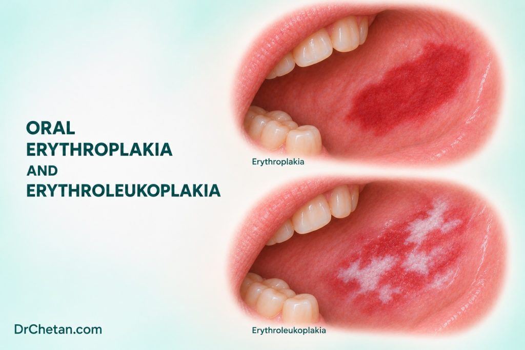
A patient has a sharply demarcated red patch on the floor of the mouth and reports little discomfort. Another has a white plaque with a red focus and palpable firmness. The two appearances raise similar questions about whether dysplasia or invasive carcinoma is already present. Redness has many causes, so the examination must establish what else could explain it and how promptly tissue assessment is needed. A persistent, unexplained red or red-white lesion with suspicious features warrants prompt biopsy by a suitably trained clinician or referral.
Start with the appearance and the differential
Oral erythroplakia is a clinical term for a red patch that cannot be characterised as another definable disease. It can have a relatively well-demarcated, velvety appearance. Erythroleukoplakia describes a mixed red and white lesion within the relevant clinical context, and is commonly treated as a nonhomogeneous leukoplakia pattern. Use “red patch” or “mixed red-white plaque” as a descriptive finding while the differential is being assessed. The clinical label does not determine whether the sample will show dysplasia, invasive carcinoma or another process.
Assess the surface under good light. Record the shape, boundary, size and location of the red and white components. Note whether a pale coating can be wiped away and whether the underlying tissue is eroded or ulcerated. Where appropriate, palpate for firmness, fixation or a deeper mass. Record bleeding or ulceration and check the remaining accessible mucosa and neck. A painless lesion may still be concerning.
| Finding | Possible explanation | How to narrow the question |
|---|---|---|
| Diffuse redness under a denture | Denture-associated inflammation or candidiasis | Look at the fit, distribution and hygiene. Assess a separate focal plaque, ulcer or firm area independently. |
| Redness with a clear contact or recent injury | Mechanical or chemical irritation | Check whether the timing and distribution fit the cause and whether the lesion resolves on an appropriate review plan. |
| Red erosion with surrounding reticular white striae | Lichenoid disease or another inflammatory disorder | Look for the wider pattern and assess a persistent or evolving focal lesion on its own. |
| Red-purple lesion suggesting a vascular process | Vascular change or another lesion with a similar colour | Describe its appearance and obtain appropriate specialist assessment before invasive sampling if vascular origin is suspected. |
| Discrete unexplained red or mixed plaque | Erythroplakia, erythroleukoplakia, dysplasia or invasive malignancy among the possibilities | Record exact location and arrange prompt assessment when the clinical features are suspicious. |
An obvious source of diffuse inflammation can coexist with a small focal lesion. For example, a broad red area that follows the outline of a denture may improve after its contributing factors are addressed. A separate indurated patch or persistent ulcer deserves its own description and decision. Where infection, trauma or inflammation is suspected, document why that explanation fits and which findings would prompt a different course.

Separate baseline cancer from later transformation
A clinical red patch can already contain carcinoma when first sampled. A systematic review of oral erythroplakia reported that approximately 43% of lesions in its initial-biopsy data were SCC at presentation. This figure came from selected observational studies, and it should not be applied to all people who present with oral redness. It illustrates why a concerning, unexplained red plaque deserves tissue assessment.
The review also analysed malignant development during follow-up. Its pooled estimate was 19.9% from three studies, with substantial heterogeneity and imprecision. This was a different outcome from carcinoma identified on the initial biopsy. The results should be kept separate in a seminar or patient discussion. Ask which lesions were followed, what their baseline diagnosis was and how long they were observed before quoting a transformation estimate.
In mixed red-white lesions, do not import the erythroplakia figure as a risk estimate for every presentation. The clinical categories, study cohorts and endpoints differ. Use the appearance to identify which regions are concerning and use clinicopathological correlation to guide care. Colour alone cannot identify the grade of epithelial change or the presence of invasion.
Refer or biopsy according to the clinical findings
Record onset and progression if known, prior treatment, relevant tobacco and areca use, alcohol exposure and whether the area is painful or bleeds. In a mixed plaque, document where the red, white, ulcerated and firm components lie. If a photo can be taken with consent, include a scale and useful landmarks. A labelled diagram can help when a lesion has several components or the photo does not show its position clearly.
For a suspicious lesion, arrange prompt biopsy or specialist referral. When a wide or heterogeneous lesion needs tissue assessment, the clinician may target the most concerning area or sample more than one region. The request should identify where each specimen came from and state whether the goal is to assess possible dysplasia, invasion or another process. Tissue depth and orientation can affect the interpretation. A prior benign result from a different part of the plaque may leave the current concern unresolved.
The NICE pathway for the UK specifically includes red or red-white oral patches consistent with erythroplakia or erythroleukoplakia. In India, route the patient through the appropriate local dental, oral medicine, oral and maxillofacial, ENT or head and neck service. State the reason for urgency and check that assessment took place. A suspicious red patch does not need to wait for an ulcer-duration threshold to be reached.
Where examination supports a low-risk, reversible explanation, a short, definite review may be appropriate after addressing it. ADA’s 2026 guide advises biopsy or referral if watchful waiting leaves an abnormality unresolved beyond 10 to 14 days without a definitive diagnosis and treatment plan. A persistent suspicious focus or new progression calls for reassessment sooner according to the findings. Light-based aids, staining or a negative cytology result cannot settle a persistent suspicious lesion.
Make urgency clear to the patient and receiving service
Explain that several conditions can cause redness and that the clinical features make further assessment important. Give the referral destination and the planned timing. A referral such as “persistent right floor-of-mouth red plaque, 13 × 8 mm, with focal induration and no convincing contact cause” communicates more than “sore mouth.” Include relevant history and the presence or absence of documented neck findings, without implying that a normal-feeling neck rules out an oral malignancy.
If the patient had prior treatment for a presumed inflammatory or infective cause, record what changed and what persisted. A wide area of irritation can improve while a focal plaque remains. A new diagnostic decision should be based on the remaining finding rather than repeating an earlier plan by default. Ensure the biopsy result or reassessment outcome is communicated and documented.
Keep a record of distinct areas
For a mixed lesion, a short map can identify a predominantly red region, a thick white region and a firm or ulcerated focus if present. Note whether these areas are contiguous and where they lie relative to teeth, the floor of mouth or a mucosal fold. Use the same landmarks on the request and any consented photographs. This lets the receiving clinician distinguish a prior sample from the current finding and choose which region warrants assessment.
Follow the report back to the mouth
When the report arrives, check that the specimen site matches the area of concern. If it shows a benign or inflammatory process, compare the expected appearance and course with the current lesion. Persistent discordance warrants discussion with the specialist and may require further assessment. If dysplasia is present, the plan depends on the grade, site, extent and overall risk assessment. Invasive carcinoma requires an oncologic workup and staging appropriate to the site.
A firm red focus within a broad plaque. An adult has an extensive white plaque with a new firm red area at one edge. A previous biopsy came from the middle of the white plaque. Map the new focus and communicate its location with the old report. Arrange prompt assessment and let the receiving clinician decide how to sample the area.
A patient with diffuse palatal redness. The redness corresponds to a denture-bearing area, with no distinct firm or ulcerated focus at that examination. If the supervising clinician finds a plausible low-risk cause, address it and arrange definite review. Reassess whether the distribution and symptoms resolve as expected. A focal residual lesion or new concerning change needs separate assessment.
The useful record links appearance to action. Describe the components, state which feature changed the level of concern, and make sure the biopsy or review outcome reaches the referring clinician and the patient.

Leave a Reply