
A patient treated for a lateral-tongue squamous cell carcinoma returns with a white patch on the opposite side. Another patient has a small visible tongue ulcer and a firm cervical node. Both cases raise questions about spread, but the mechanisms can differ. The contralateral patch may be an unrelated disorder, a potentially malignant change or a new tumour. The neck node may contain cells from the original primary. Understanding the difference between a susceptible epithelial field, local invasion and metastasis helps dental students document the finding and interpret the next report.
Changes accumulate in the epithelium
Oral squamous cell carcinoma arises from epithelium with squamous differentiation. Long-term exposure to tobacco, areca nut or alcohol can contribute to molecular damage and selection of altered cell populations. A clone is a population descended from an ancestral cell. Clones can acquire different alterations as they expand, and several clones can coexist within a region of epithelium. Their growth also depends on the surrounding tissue, immune response and additional changes. The result is a branching process with several possible outcomes.
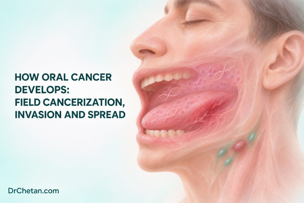
TP53 and CDKN2A are useful examples of genes involved in cell-cycle control. Loss of normal p53 function can impair responses to DNA damage. Changes that affect CDKN2A can disrupt restraints on cell-cycle progression. The Cancer Genome Atlas documented diverse alterations across head and neck SCCs, with differences by site and HPV association. These genes illustrate mechanisms. There is no routine chairside test based on them that can decide which oral patch will become invasive.
What an altered field explains
Field cancerization describes a region of epithelium in which carcinogenic exposure and expansion of altered cells can create conditions for more than one lesion or tumour. Some molecular studies have found that separated lesions share alterations, consistent with a common ancestral clone. Other lesions can arise independently in the same exposed mucosa. A clinical examination shows the visible and palpable features. It cannot identify the borders or ancestry of every altered clone.
This helps explain why follow-up includes the treated site and the rest of the mouth. A clear surgical margin means that the pathologist found no qualifying tumour at the assessed margin under the reporting protocol. It is a key local finding. It cannot quantify future risk throughout the oral mucosa. Conversely, a new plaque or ulcer after treatment is not automatically a recurrence. Record its relationship to the previous site, compare with earlier descriptions and arrange assessment based on the findings.
Separate an OPMD, dysplasia and invasive cancer
An oral potentially malignant disorder, or OPMD, is a clinical risk category. Leukoplakia and oral submucous fibrosis are examples. Epithelial dysplasia is a histological finding involving abnormal cellular and architectural features within the epithelium. A clinical lesion can contain areas with different histology. Its behaviour depends on the condition, site, changes over time and whether the tissue sampled represents the area of concern. Many lesions never progress, and some invasive cancers present without a recognised preceding disorder.
Invasion occurs when malignant epithelial cells extend into the supporting tissue. The pathologist assesses the relationship between epithelium and stroma, the pattern of invasive nests or cells, and features that support interpretation. A biopsy with an appropriate epithelial-stromal interface helps answer whether invasion is present. A surface fragment may contain abnormal epithelium without including the invasive component. If the tissue result and clinical concern do not align, reassess the site and adequacy with the treating team.
Consider the surrounding tissue
The stroma around invading tumour cells contains matrix, fibroblasts, vessels and immune cells. The interaction between tumour and surrounding tissue can influence growth and routes of invasion. In practice, the student sees the consequences through clinical examination, imaging and pathology. An indurated margin or restricted movement may indicate deeper involvement, but inflammation and scarring can produce overlapping findings. Record what was observed and let the full workup establish extent.
Dysplasia grade, depth of invasion and tumour differentiation describe different things. Dysplasia grade describes changes in epithelium. Depth of invasion is a measurement of an invasive tumour. Differentiation describes how closely the tumour retains features of squamous maturation. A report can contain more than one of these observations. Identify which tissue and specimen the report describes before using any feature to discuss risk or extent.
Follow the three routes of spread
| Route | What happens | What the care team evaluates |
|---|---|---|
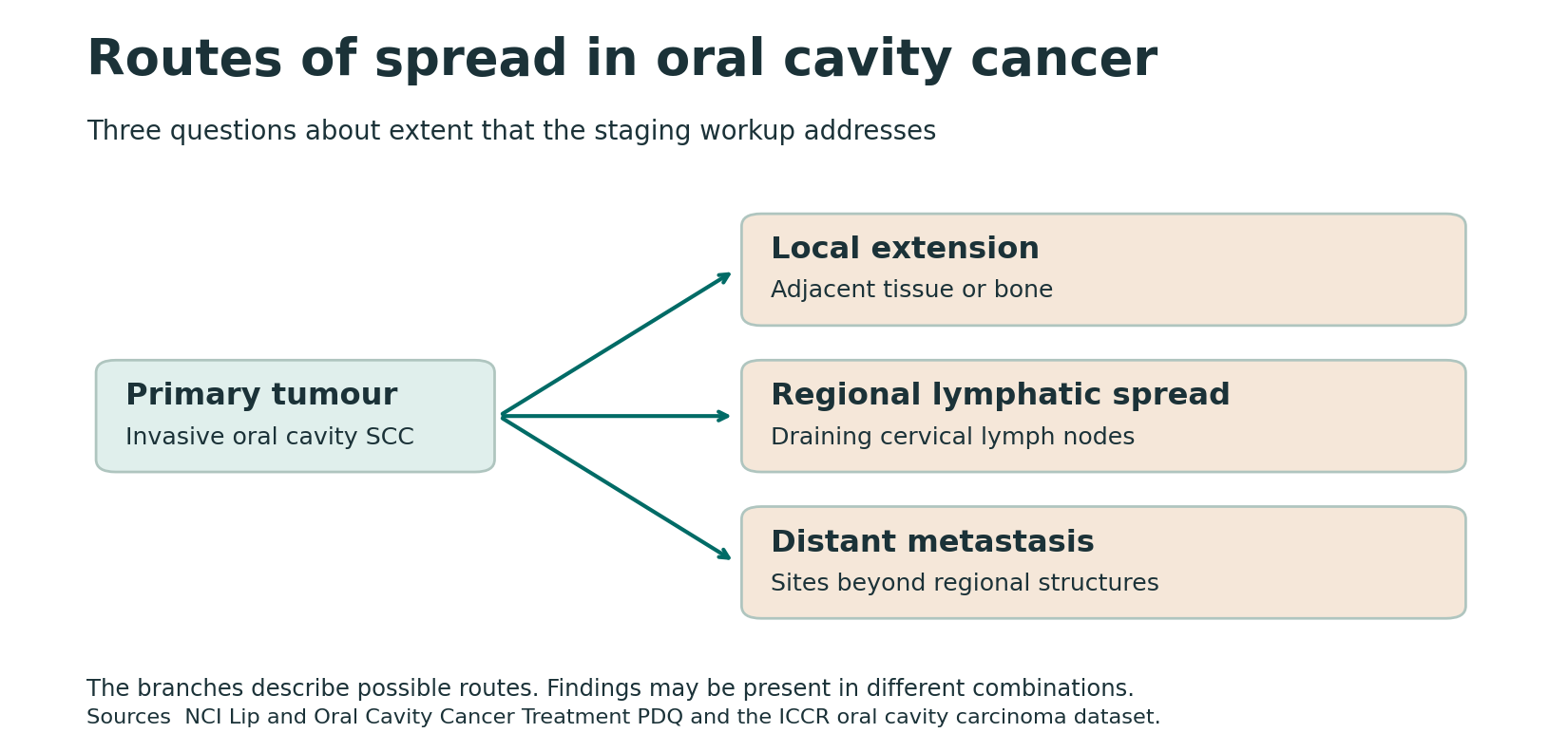
| Local extension | Invasive tumour grows into contiguous soft tissue, muscle or bone | The primary site, depth of invasion, involved structures, resection margins and function. |
| Regional lymphatic spread | Tumour reaches lymph nodes draining the primary region | Nodal side and levels, clinical and imaging findings, pathology and extranodal extension when present. |
| Distant metastasis | Tumour establishes deposits beyond regional structures | Staging investigations chosen for the clinical setting and whether a distant lesion is metastatic, a new primary or another process. |
The apparent surface dimensions of a lesion can underestimate its deeper extent. In the oral cavity SCC staging framework, depth of invasion is measured histologically from a reconstructed level of adjacent normal mucosal basement membrane to the deepest invasive point. Tumour thickness measures from the tumour surface and can differ in exophytic or ulcerated lesions. Adjacent structure involvement adds separate information. For example, a gingival primary near bone and a lesion that has invaded through qualifying bony structures do not carry the same implications. Apply the specific chapter criteria to the final clinical and pathological findings.
Cells can enter lymphatic channels and reach regional cervical nodes. A clinically node-negative neck can still contain microscopic metastases. The likelihood and management depend on the site, stage and other findings. Midline proximity can affect drainage and specialist assessment of the opposite neck. Extranodal extension means that a nodal metastasis extends beyond the nodal capsule into surrounding tissue. The nodal report matters separately from the report on the primary tumour.
Distant metastasis involves additional steps, including survival during transport and growth in another tissue. A distant lesion on imaging can have several explanations. A new lung finding, for example, may prompt workup for a metastasis, a separate lung primary or a benign condition. The oncology team uses the pattern, timing, imaging and tissue when available to resolve the question. A spread diagram represents possible routes, and an individual tumour can present with one route evident while others are absent or undetected.
Read the pathology report as a set of observations
A diagnosis of invasive SCC identifies the process, and the resection report adds detail about its extent. Along with the site and dimensions, review depth of invasion, margins, perineural invasion and lymphovascular invasion where applicable. Perineural invasion describes a relationship between tumour and nerves. Lymphovascular invasion describes tumour within vascular spaces under pathological criteria. These findings inform postoperative risk assessment. Neither finding by itself establishes that the patient has a distant metastasis.
The neck report can identify involved nodes and extranodal extension. Review it alongside the primary resection. Clinical staging describes the evidence available before definitive treatment, and pathological staging can add information from resection and nodal assessment. A pathology report confirming local invasion answers a tissue question. To establish the overall stage, combine the primary tumour, regional node and distant assessments within the appropriate staging system.
Distinguish recurrence, a second primary and metastasis
Local recurrence is return of disease at or near the treated primary site. Regional recurrence involves the regional drainage basin. A second primary is a separate tumour, classified using clinical and pathological criteria and sometimes further molecular comparison. Metastasis is spread of tumour from a primary site to another location. The distance from a prior tumour and the elapsed time can inform the workup. The team needs the new lesion’s site, histology and relationship to the original disease before settling the classification.
Consider the patient with the opposite-side plaque. Document its exact subsite, dimensions, surface and any induration or ulceration. Compare the finding with the earlier oral chart, discuss relevant exposures and contact the oncology or oral medicine team when assessment is warranted. A case summary should keep the history of treated SCC and the observed new finding as separate facts until there is a diagnosis.
For the patient with the tongue ulcer and cervical lump, record the ulcer’s location and extent and the side and location of the lump. Prompt assessment may involve biopsy of the oral lesion, imaging and specialist-directed nodal sampling. The physical findings can suggest a relationship. Pathology and the staging workup determine how the two findings fit together.
At follow-up, compare the original tumour bed, the rest of the oral mucosa and the neck with prior findings. Ask about progressive symptoms and barriers to attending reassessment. A field model explains why the clinician keeps looking beyond the original lesion. The practical decision for any new abnormality still comes from its clinical features, history and diagnostic assessment.

Leave a Reply