
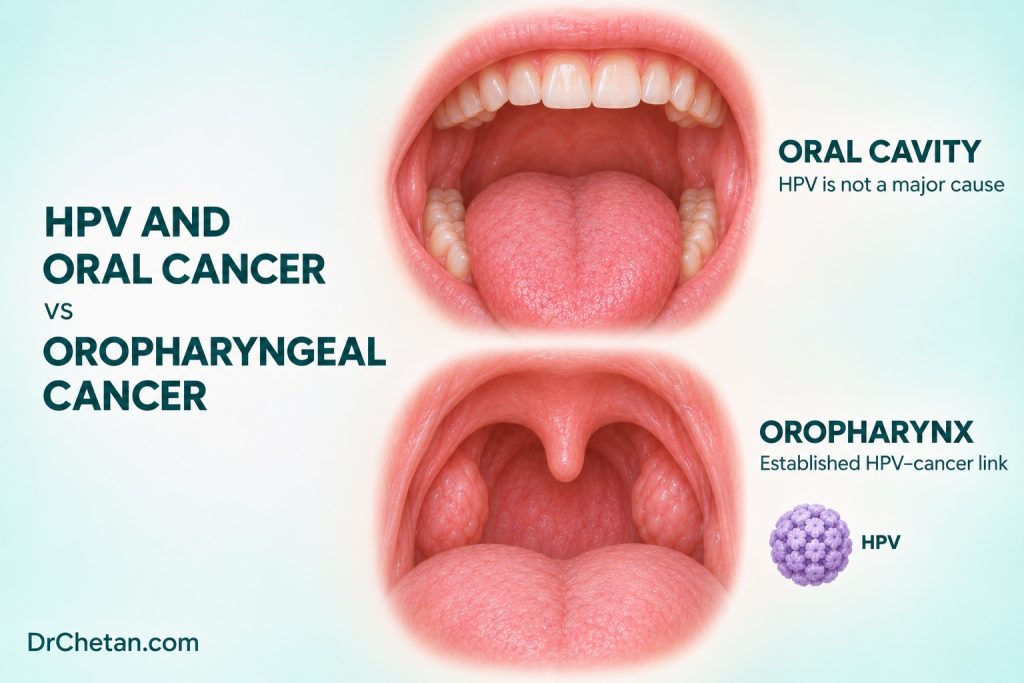
“HPV-positive tongue cancer” leaves several questions unanswered. Is the tumour on the mobile tongue or at the tongue base? Was p16 immunohistochemistry performed on tissue, or was an HPV-specific test performed on a node sample? A clinician needs those details before applying staging or discussing prognosis. High-risk human papillomavirus (HR-HPV) causes a subset of oropharyngeal squamous cell carcinomas, particularly in the palatine and lingual tonsillar regions. The same association and testing rules do not apply routinely to a confirmed oral cavity squamous cell carcinoma.
Establish the site before interpreting the test
The mobile tongue belongs to the oral cavity. The base of tongue, which includes lingual tonsillar tissue, belongs to the oropharynx. Palatine tonsils, tonsillar pillars, soft palate and oropharyngeal walls are also in the oropharynx. A tumour arising on the lateral mobile tongue can extend toward the base. A small tonsillar primary can be hard to see and first present as a cervical nodal mass. Give the exact site or state which part of the lesion cannot be localised. The receiving team can correlate the examination with endoscopy, imaging and pathology.
A useful referral distinguishes where symptoms are felt from where a lesion is observed. “Left lateral mobile tongue with posterior extent incompletely visible” directs attention to the visible lesion and preserves uncertainty. “Persistent unilateral cervical lump with no primary visible on dental examination” records a different presentation. It allows the specialist to consider an occult head and neck primary alongside other causes of a neck mass.
How high-risk HPV drives some oropharyngeal cancers
HPV infection is common, and most infections clear. Persistent high-risk infection can contribute to cellular transformation. In an HPV-driven tumour, viral E6 and E7 proteins interfere with important cell-cycle controls. E6 promotes loss of p53 function, and E7 disrupts the retinoblastoma pathway. The resulting increase in p16 expression can be detected by immunohistochemistry. This is one reason p16 can serve as a surrogate in the appropriate tumour and specimen.
An HPV-specific assay asks a different question from p16 immunohistochemistry. p16 staining measures a host-cell protein. RNA in situ hybridisation can show expression of viral transcripts in the tissue. A validated DNA PCR assay detects viral DNA and requires interpretation within its intended diagnostic setting. CAP calls for HPV-specific assays with appropriate performance and coverage of high-risk types beyond HPV16. Detection of viral material and attribution of an individual tumour to HPV must be interpreted with site, morphology, specimen type and the test performed.
Nonkeratinising morphology can support consideration of HPV-associated oropharyngeal SCC, but pathology still follows the testing recommendations. Keratinisation and p16 can be discordant. A visible oral lesion or an image of a throat finding cannot establish whether a tumour is HPV-associated.
Which specimen and which test
| Clinical and specimen setting | How CAP approaches testing |
|---|---|
| Newly diagnosed oropharyngeal SCC, suitable tissue | Assess HR-HPV status. p16 immunohistochemistry is the usual surrogate in the guideline’s defined non-cytology setting. |
| Oropharyngeal tissue with equivocal p16, discordant morphology or other specified caveat | Perform HPV-specific testing. Examples include selected nontonsillar sites, large overlapping lesions and low-prevalence geographic settings. |
| Metastatic cervical SCC with unknown primary, non-cytology tissue | Perform HPV-specific testing or use p16 immunohistochemistry followed by HPV-specific testing for p16-positive tumours. |
| Cervical node fine-needle aspiration in the setting specified by CAP | Prefer HPV-specific testing. CAP describes selected exceptions and advises testing available tissue when FNA HPV testing is negative. |
| Confirmed primary oral cavity SCC | Routine HR-HPV testing for prognostic purposes falls outside CAP’s recommendation for this site. A particular diagnostic question can justify specialist-directed testing. |
For tissue in which p16 immunohistochemistry is indicated, the CAP summary defines positivity as at least 70% nuclear and cytoplasmic staining with at least moderate-to-strong intensity. This scoring rule applies to the specified tissue setting. Fine-needle aspiration material has its own recommendations and technical limitations. In tissue cases, CAP specifies HPV-specific testing when p16 is equivocal, the stain and morphology disagree, or other caveats apply. A reported percentage therefore belongs with the specimen, staining intensity, interpretation and site.
When a node is the first specimen, the main question is where the tumour started. High-risk HPV testing can help direct the search for an oropharyngeal primary in metastatic cervical SCC. The team still correlates the result with nodal location, imaging, endoscopy and other pathology. A negative FNA result does not settle the question if further tissue becomes available and the clinical suspicion remains.
Translate a laboratory report into plain language
For a suitable tonsillar resection, a report might state that p16 showed diffuse, strong nuclear and cytoplasmic staining consistent with positivity in that setting. For a different case it might report a positive high-risk HPV RNA in situ hybridisation result. The first describes a host marker that serves as a surrogate when the criteria apply. The second uses a viral target. Both need a site and specimen description. When reading the report, check the actual test line and interpretation rather than shortening either result to “HPV positive” without context.
An equivocal or discordant report is a reason to communicate with the pathology team. Ask what tissue was sampled, whether the tumour is clearly from an oropharyngeal site and whether a confirmatory assay is needed or pending. Older reports may describe an eighth-edition p16-positive staging group. If presenting a historical case alongside a current case, name each system and date. This preserves how the stage was assigned and prevents a comparison of categories that do not follow identical rules.
Staging and prognosis are separate decisions
As of the AJCC 2026 staging table, HPV-associated oropharyngeal carcinoma is staged using Version 9 for diagnoses from 1 January 2026 under that implementation. The oral cavity chapter remains in the eighth edition. The appropriate scheme depends on tumour site, histology, the evidence for HPV association and diagnosis date. When tests disagree or the primary site remains uncertain, the treating team reconciles the data with the applicable pathology and staging protocol. A positive p16 result on its own should never cause a confirmed mobile-tongue primary to be staged as an oropharyngeal cancer.
In clinical cohorts, patients with HPV-associated oropharyngeal SCC generally have better outcomes than those with HPV-independent oropharyngeal SCC. Individual prognosis still depends on disease extent, tobacco history, health, treatment and other factors. A survival figure from a particular study reflects its patients, therapies and endpoints. It cannot be transferred to a p16-positive oral cavity lesion or used to select less intensive treatment in a dental setting. Treatment decisions belong to the oncology team.
Explain prevention without implying a diagnosis
HPV vaccination helps prevent new infections with covered types, including high-risk types associated with oropharyngeal cancer. It does not treat an existing infection or tumour. Eligibility and schedules depend on the current local programme and individual clinical circumstances. A discussion about vaccination is separate from diagnostic workup for a suspicious lesion or neck lump. Routine testing for oral HPV in people without symptoms is not a substitute for investigating a suspicious clinical finding.
Avoid assumptions about an individual’s relationships, exposures or when an infection was acquired. An HPV-associated tumour can emerge long after the initial infection, and a pathology report cannot establish when or from whom a person acquired it. A useful conversation explains what the test means for this tumour and directs personal questions to the treating team. Continue to address tobacco and areca use when relevant. They remain important prevention topics regardless of an oropharyngeal tumour’s HPV status.
Two cases to distinguish at chairside
A confirmed mobile-tongue primary with a positive p16 result. A pathology report mentions p16 staining in a lesion documented on the right lateral mobile tongue. Ask the team what question prompted the stain and confirm the site on the referral and imaging. If this is an oral cavity primary, the oral cavity pathway applies. A laboratory marker alone does not relocate the primary.
A neck mass with no primary visible during dental examination. A patient has a persistent unilateral neck lump and pain on swallowing. Their oral cavity examination does not reveal a cause. Arrange prompt specialist assessment and document the examination limits. If needle sampling identifies metastatic SCC, the specialist team can pursue the specimen-specific HPV workup and investigate an occult primary. The first dental decision is referral and documentation.
For a case presentation, report four details together: the anatomical primary site or uncertainty, the sample type, the HPV or p16 test and its interpretation, and the staging protocol used. This makes clear which parts of the conclusion come from examination, which from the laboratory and which require further workup.

Leave a Reply