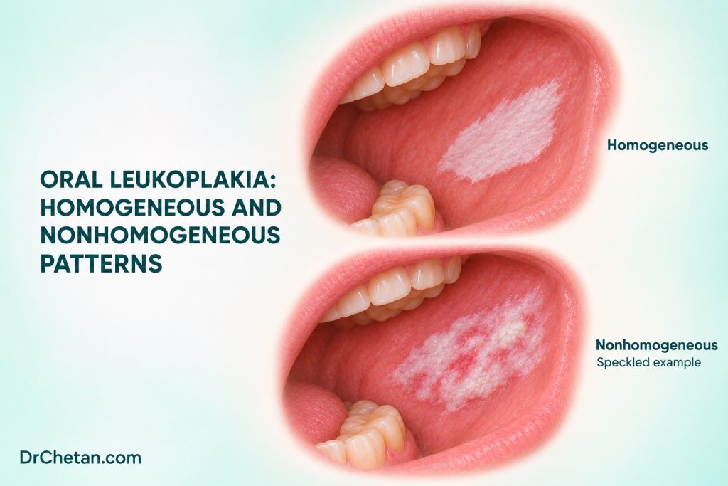
Two white plaques can look similar at a glance. One is flat and uniformly white. The other has a small red area, a raised nodule and a firm margin. The surface pattern helps determine how urgently to investigate and which areas may need sampling. The tissue diagnosis still requires clinicopathological correlation. For dental students, the sequence is to describe the plaque, consider specific explanations, assess suspicion and make a plan that follows the patient through biopsy or reassessment.
Establish what the clinical label means
Oral leukoplakia is a clinical diagnosis of exclusion for a predominantly white plaque of uncertain cancer risk after other definable disorders have been excluded. The description “white patch” is the start of the examination. A firmly attached white area may reflect increased keratin, epithelial change or another process. Its colour alone cannot establish whether there is epithelial dysplasia or invasive carcinoma. A plaque can contain regions with different findings, so the final interpretation depends on which part was examined and sampled.
The clinical label, the microscopic diagnosis and the risk assessment describe different things. A report of hyperkeratosis with no dysplasia identifies the sampled tissue. A report of dysplasia describes an architectural and cellular abnormality in that sample. A diagnosis of invasive SCC means malignant epithelial cells have entered the underlying tissue. If histology and clinical findings do not fit, revisit the site and representativeness of the sample.
Recognise homogeneous and nonhomogeneous patterns
| Pattern | Typical description | What to assess |
|---|---|---|
| Homogeneous | Predominantly white and relatively uniform in colour and surface, often flat or slightly raised | Dimensions, margins, subsite, firmness and change compared with earlier records. |
| Red and white | Red areas interspersed with or adjacent to white change, sometimes termed erythroleukoplakia | Where the red change is most distinct and whether there is ulceration, nodularity or induration. |
| Nodular or irregular | Raised foci or an uneven surface within a plaque | Which focus is new or firm and whether the plaque is changing. |
| Verrucous | Rough, papillary or warty surface | Whether the finding is focal or part of progressive multifocal disease, and which areas need specialist sampling. |
Homogeneous plaques generally have a lower observed transformation risk than nonhomogeneous plaques in study cohorts. Homogeneity alone cannot guarantee the absence of dysplasia or cancer. Risk assessment also considers site, size, persistence, histology and whether there is a new symptom or change. Use the pattern to describe what is present today. Keep the dated record so a later examiner can tell which features have changed.
Check common explanations for a white plaque

Frictional change. A rough area in contact with an obvious mechanical source can support a reactive explanation if the location and time course fit. Record the source and clinical appearance. Where findings are suitable for short observation, address the irritation and arrange reassessment. A progressive plaque, unexplained red component or firm focus warrants assessment on its own merits.
Candidiasis. Pseudomembranous candidiasis often has material that can be wiped away, with an erythematous surface beneath. Some forms, including chronic hyperplastic candidiasis, can present as an adherent white lesion. Clinical context and response to appropriate treatment can inform the differential, and a persistent or suspicious plaque may require tissue assessment. Avoid using a positive fungal finding to explain away a separate focal abnormality.
Lichenoid disease. Bilateral reticular striae and a characteristic distribution can suggest oral lichen planus or a lichenoid disorder. A focal plaque or erosion may need further assessment when it is asymmetric, evolving or discordant with the background pattern. Record the whole-mouth distribution and the specific area that prompted concern.
Oral hairy leukoplakia. This is a separate Epstein–Barr virus-associated condition, often seen as a corrugated adherent plaque along the lateral tongue, especially in the setting of immunosuppression. The shared word leukoplakia does not make the conditions equivalent. Review the clinical context and refer for confirmation when the appearance or history is uncertain. A focal red, ulcerated or firm area still needs its own assessment.
Record the lesion and identify the important biopsy question
Ask about onset, changes, prior similar lesions and prior biopsy sites. Record tobacco, areca and alcohol separately. Examine both sides of the mouth and the neck as appropriate. For each lesion, give the exact subsite, side, greatest visible dimensions, whether the lesion wipes away, its colour and texture, and any firmness or ulceration. A consented dated photograph with scale and landmarks can support a written map.
If the lesion is suspicious, arrange prompt biopsy within the clinician’s competence or refer to a service that can assess it. When a wide plaque has different components, a clinician may need samples from more than one site. The request should say which sample came from the red area, raised focus or other concerning region. If the visible and palpable abnormality is more extensive than the specimen, say so. A superficial or unrepresentative sample can leave invasion unresolved.
When there is a convincing low-risk explanation, a short reassessment can be reasonable after addressing it. ADA’s 2026 chairside guide advises biopsy or referral if a finding managed by watchful waiting persists beyond 10 to 14 days without a definitive diagnosis and treatment plan. A suspicious lesion warrants prompt assessment at the initial visit. Cytology has a limited contingency role under the guideline. A negative adjunctive result cannot settle a suspicious persistent plaque.
Compare the report with the clinical photograph and map
When the report arrives, locate its sample on the original map. A nonhomogeneous plaque can be divided into regions by landmarks, for example a flat white area posterior to a molar and a raised red-white focus closer to the floor of mouth. If the report describes a bland keratotic sample from the first region, ask whether the second region was assessed or remains a diagnostic question. The written correlation helps prevent a reassuring result from being applied to tissue that was never sampled.
Also review how the clinical label was reached. A pathology report may give an alternative explanation, such as inflammation or infection. Compare that explanation with the site, appearance and subsequent course. When the report and examination remain discordant, discuss the discrepancy with the specialist and document the plan for reassessment or further sampling. The aim is to resolve the mismatch and preserve the details needed for follow-up.
Discuss risk without turning a pooled estimate into a prediction
A systematic review of 55 studies reported a pooled malignant transformation proportion of 6.64%, with a 95% confidence interval of 5.21% to 8.21%. Between-study heterogeneity was high. Nonhomogeneous appearance, greater size, lateral tongue location and epithelial dysplasia were associated with higher risk across the included studies. The pooled proportion combines cohorts with different case definitions, treatment and follow-up periods. It cannot tell a patient the probability that their plaque will become cancer in the next year.
Specialists consider observation, excision or other management according to clinical features, histology, extent, morbidity and capacity for follow-up. A randomised trial in selected nondysplastic lesions informs observation in that setting. It cannot be generalised to lesions with dysplasia or a discordant suspicious examination. For extensive or heterogeneous plaques, repeat or multi-site sampling may help resolve a management decision. Any plan should include review of the treated site and the rest of the mouth.
Two decisions to practise
A uniform plaque on the buccal mucosa. A patient has a thin, uniformly white plaque next to a fractured restoration. It is soft on palpation and there is a plausible contact pattern. If the supervising clinician judges short review appropriate, record the baseline and address the contact. At the booked visit, compare the site, surface and size. Persistence without a clear diagnosis calls for further assessment.
A red-white plaque with a firm nodule. Another patient has a nonhomogeneous plaque on the ventrolateral tongue with a firm red focus that was not on the previous chart. Record the new finding and arrange prompt assessment. Give the specialist the previous report and its sample location. A prior result from a flat white region and the current firm red focus describe different parts of the clinical problem.
After the report returns, reconcile it with the examination and document who will follow the patient. Advise earlier reassessment if the plaque grows, develops a red or firm component or ulcerates. A usable plan gives the patient a destination, a review date and a clear way to report change.

Leave a Reply