An oral histology and embryology guide for dental students, covering the dental lamina, bud, cap, and bell stages, tissue origins, dentin and enamel formation, and the beginning of root development.
A tooth does not begin as a miniature mineralised crown. It begins through interactions between oral epithelial cells and the ectomesenchyme beneath them. Those interactions establish a tooth germ, shape its future crown, generate specialised cells, and organise the deposition of daental tissues.
The familiar bud, cap, and bell stages describe the changing appearance of the developing enamel organ. They are useful landmarks within a continuous process, rather than three isolated events that explain everything about tooth formation.
For a dental student, understanding tooth development becomes much easier when you keep three questions separate: What does the tooth germ look like? Which cells are differentiating? What tissue is being formed? This guide connects those questions and provides diagrams, comparison tables, and revision questions.
In this guide:
- First distinguish stages from processes
- Where do the tissues of a tooth come from?
- Initiation and the dental lamina
- The bud stage
- The cap stage
- The bell stage
- Histodifferentiation versus morphodifferentiation
- Advanced bell stage and the start of hard-tissue formation
- Dentinogenesis and amelogenesis: compare the cells and direction
- Apposition, mineralisation, and maturation
- Reduced enamel epithelium and preparation for eruption
- How root development begins
- Primary teeth, permanent successors, and permanent molars
- Epithelial remnants: Serres versus Malassez
- How to recognise developmental stages in a slide
- Why these stages matter clinically
- A simple revision method
- Practice questions
- Final revision checklist
First distinguish stages from processes
You will encounter both a morphological classification and a list of physiological processes. They describe different aspects of the same developing tooth.
| Classification | What it describes | Examples |
|---|---|---|
| Morphological stages | The changing shape and organisation of the enamel organ and tooth germ | Bud, cap, early bell, late bell |
| Developmental processes | What cells and tissues are doing | Initiation, proliferation, histodifferentiation, morphodifferentiation, apposition, mineralisation and maturation |
Histodifferentiation means cells acquiring specialised identities, such as odontoblasts and ameloblasts. Morphodifferentiation means the developing tooth acquiring its characteristic form. Apposition means matrix being deposited in layers.
These processes overlap. Proliferation contributes to more than one morphological stage; crown shaping is a progressive activity; and matrix deposition begins in the advanced bell stage rather than waiting for a completely unrelated developmental episode. A list of phases is a learning framework, not a set of watertight compartments.
Where do the tissues of a tooth come from?
Tooth development depends on epithelial–ectomesenchymal interactions. The dental epithelium comes from oral ectoderm. The relevant craniofacial ectomesenchyme is largely derived from migrating neural crest cells.
This distinction matters because enamel and dentin have different cellular origins. Enamel is made by epithelial-derived ameloblasts. Dentin is made by odontoblasts derived from the dental papilla’s ectomesenchyme. Do not label the papilla as part of the enamel epithelium merely because the two lie next to each other.
As the tooth germ becomes organised, recognise its three principal components:
| Component | Basic origin and position | Main contribution |
|---|---|---|
| Enamel organ | Epithelial component, shaped around the dental papilla | Its inner enamel epithelial lineage gives rise to ameloblasts that form enamel; other parts support development or have later epithelial roles |
| Dental papilla | Condensed ectomesenchyme within the concavity of the enamel organ | Peripheral odontoblasts form dentin; the remaining central tissue develops into the pulp |
| Dental follicle, or dental sac | Condensed ectomesenchyme surrounding the enamel organ and papilla | Contributes to cementum, periodontal ligament, and the tooth’s supporting alveolar tissues |
The phrase “enamel organ forms enamel” is convenient shorthand. It does not mean that every cell in the enamel organ turns into enamel. Likewise, the dental papilla is not simply a store of future dentin: it also supplies the future pulp.
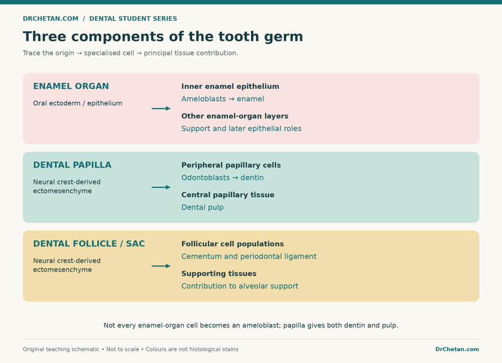
Figure 1. Follow the cell lineage, not just the name of the component. The diagram shows the principal undergraduate tissue-derivative relationships.
Initiation and the dental lamina
In a conventional account of early primary-tooth development, the first epithelial changes are described around the sixth week of intrauterine life. Local epithelial thickening and growth into the underlying ectomesenchyme establish the dental lamina along the future dental arches. Individual tooth-forming sites develop along it.
The dental lamina is an epithelial structure, not a mineralised plate. It provides the early epithelial connection between the developing tooth germs and the oral epithelium.
A related epithelial structure, the vestibular lamina, is associated with formation of the oral vestibule between the lips or cheeks and the tooth-bearing part of the jaws. Keep the two names separate: dental lamina relates to tooth development; vestibular lamina relates to the vestibular space.
Do all teeth begin developing at the same time?
No. Different teeth start and progress at different times. The primary dentition begins developing prenatally, while the permanent dentition follows a staggered programme that extends across prenatal and postnatal life.
For example, a tooth germ can be in an early shape stage while another is already depositing matrix. A single age assigned to “the cap stage” cannot be applied to every tooth in the mouth. Learn the developmental sequence first, then use the tooth-specific chronology required by your prescribed text.
The bud stage
The bud stage is characterised by a rounded or ovoid epithelial outgrowth extending from the dental lamina into the underlying ectomesenchyme. Budding of the primary teeth is commonly described around the eighth prenatal week in introductory accounts.
At this point, proliferation is prominent, but the epithelial component has not yet acquired the deep concavity or the clearly organised four-layer arrangement seen later. The ectomesenchyme around the growing bud becomes condensed.
What should you recognise in a bud-stage section?
- The surface oral epithelium.
- The epithelial connection or dental lamina.
- The rounded epithelial tooth bud.
- The surrounding, increasingly condensed ectomesenchyme.
There is no mature enamel cap, completed dentin, or finished pulp chamber in this early bud. The name describes the epithelial shape, not a miniature erupted tooth.
The underlying papillary and surrounding follicular regions become increasingly distinct as development advances. In a practical slide, use the overall arrangement rather than assuming that every early component will be sharply outlined.
The cap stage
Unequal growth produces an indentation on the deep surface of the epithelial bud. The enamel organ now resembles a cap over the condensed dental papilla. The surrounding dental follicle is more clearly recognisable.
The major spatial relationships are the important part: enamel organ outside and above the papillary concavity, papilla within that concavity, and follicle around the tooth germ. These are interacting tissues, not empty spaces inside a shell.
Within the cap-stage enamel organ, identify:
- Outer enamel epithelium: the epithelial boundary towards the follicle.
- Inner enamel epithelium: the epithelial boundary facing the papilla.
- Developing stellate reticulum: the central epithelial region whose cells become separated by fluid-rich extracellular spaces and develop a star-like appearance.
The stratum intermedium becomes a distinct additional layer in the bell stage. This is a useful comparison when moving between cap- and bell-stage descriptions.
What does the enamel knot do?
The enamel knot is a transient epithelial signalling centre involved in organising tooth morphogenesis. It influences the behaviour of neighbouring cells and contributes to the patterning of the developing crown. In multicusped teeth, secondary enamel-knot signalling centres are associated with future cusp positions.
Do not describe the enamel knot as a mineralised piece of enamel or as a cusp that simply grows larger. Its significance is primarily organisational and signalling-related, and its activity is temporary.
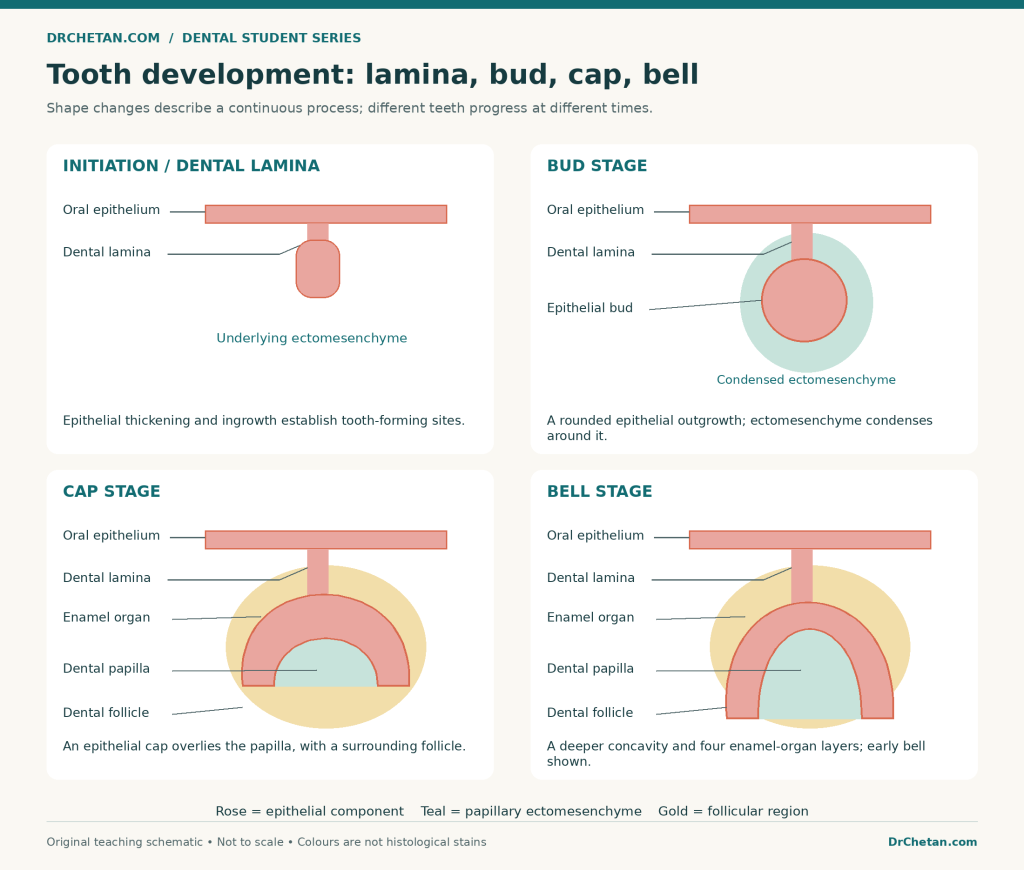
Figure 2. Schematic sections showing shape changes. Colours distinguish epithelial tissue, papilla, and follicle; they do not represent routine histological stain colours. Stage sizes and timings are not drawn to scale.
The bell stage
The epithelial concavity deepens and the enamel organ becomes more bell-shaped. At the same time, the cells and tissues become more specialised and the future crown pattern becomes more defined.
This is why the bell stage is associated with both histodifferentiation and morphodifferentiation. It is not simply a cap stage made larger. Early and late bell stages are useful subdivisions, especially when discussing the onset of dentin and enamel production.
The four layers of the bell-stage enamel organ
| Layer | Where to find it | Main point to remember |
|---|---|---|
| Outer enamel epithelium, OEE | Outer boundary facing the dental follicle | Maintains the outer epithelial boundary and participates in exchange with the surrounding environment |
| Stellate reticulum, SR | Loose central region between the outer epithelium and the deeper layers | Star-shaped epithelial cells and fluid-rich spaces provide structural support |
| Stratum intermedium, SI | Between the stellate reticulum and inner enamel epithelium | Closely associated with ameloblast function and enamel formation, including metabolic and mineralisation support |
| Inner enamel epithelium, IEE | Inner boundary facing the dental papilla | Contributes to crown form, induces papillary cell differentiation, and gives rise to the ameloblast lineage |
The stratum intermedium lies next to the inner enamel epithelium, not between the outer enamel epithelium and the follicle. Students often remember all four names but place this layer on the wrong side of a diagram.
The enamel organ is avascular; it is supported through exchange with surrounding vascular connective tissues. Spaces in the stellate reticulum should not be mistaken for a collection of blood vessels.
What is the cervical loop?
The cervical loop is the margin where the inner and outer enamel epithelia meet. This is an epithelial junction, not the future root canal. Its later development is important because the inner and outer epithelial layers extend to form Hertwig’s epithelial root sheath.
What lies between the IEE and the papilla?
Before hard-tissue formation, the epithelial and ectomesenchymal compartments are separated by a basement membrane. Their interface helps establish the location of the future dentinoenamel junction, or DEJ.
Do not say that the basement membrane simply becomes a thick layer of enamel or dentin. The relationship changes as the adjacent cells differentiate and matrix is deposited. The future DEJ is the interface between the developing enamel and dentin.
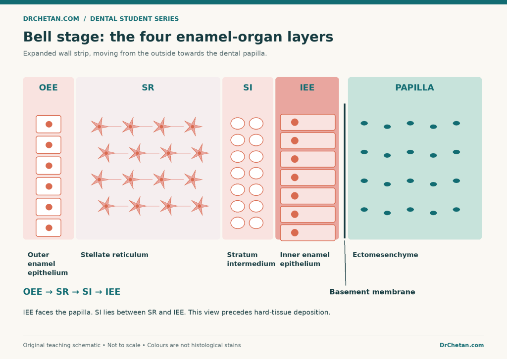
Figure 3. Expanded schematic of the tissue order on one side of a bell-stage enamel organ before hard tissues are laid down. Widths are exaggerated for readability; this is not a micrograph.
Histodifferentiation versus morphodifferentiation
These terms are connected, but they answer different questions.
Histodifferentiation asks: what kind of cell is this becoming? The odontoblast acquires the structure and activity needed to produce dentin matrix. The ameloblast lineage acquires its specialised enamel-forming role.
Morphodifferentiation asks: what form is the developing tooth acquiring? Folding and growth of the enamel epithelium contribute to the crown outline and cusp pattern. Tooth shape emerges through organised tissue interactions, not through a finished tooth being carved out of an undifferentiated block.
In a viva, give one example of each. “Odontoblast differentiation” illustrates cell specialisation. “Establishment of the crown’s cusp pattern” illustrates morphological development. Avoid defining both terms as simply “the tooth develops.”
Advanced bell stage and the start of hard-tissue formation
The central sequence to understand is reciprocal induction between the epithelial and papillary cell populations. The cells do not work independently, and the developmental history of one tissue affects the other.
Step 1: The inner enamel epithelial cells become preameloblasts
Cells in the IEE elongate and specialise. Through epithelial-ectomesenchymal interactions, the developing preameloblast population influences adjacent peripheral papillary cells.
Step 2: Peripheral papillary cells differentiate into odontoblasts
The peripheral dental papilla cells adjacent to the future DEJ become odontoblasts. These are ectomesenchymal-derived cells, even though their differentiation is influenced by the neighbouring epithelium.
Step 3: Odontoblasts deposit predentin and dentin formation begins
Odontoblasts secrete predentin, the initially unmineralised dentin matrix. Mineralisation follows to produce the first dentin. Crown hard-tissue formation begins in the future cusp-tip or incisal regions and extends towards the cervical part of the crown.
Step 4: Secretory ameloblasts begin enamel matrix deposition
Formation of the initial dentin is followed by completion of the adjacent ameloblasts’ secretory differentiation and the beginning of enamel matrix secretion.
Therefore, the first dentin is formed before the first enamel at the developing crown interface. This does not mean epithelial differentiation starts only after odontoblasts appear: preameloblast development and signalling are already involved earlier. Distinguish the start of cell differentiation from the order of hard-tissue deposition.
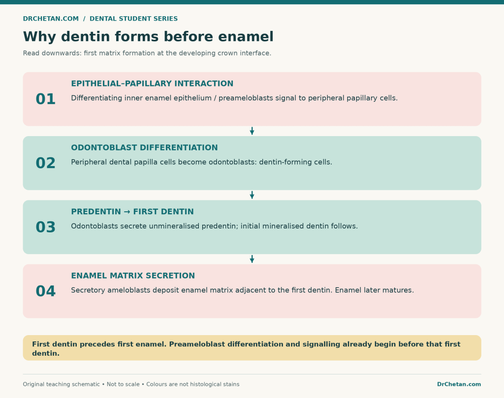
Figure 4. First dentin precedes first enamel. The diagram separates cell differentiation, matrix secretion, and mineralisation rather than treating them as one event.
Dentinogenesis and amelogenesis: compare the cells and direction
| Feature | Dentin formation | Enamel formation |
|---|---|---|
| Forming cell | Odontoblast | Ameloblast |
| Developmental lineage | Peripheral dental papilla ectomesenchyme | Inner enamel epithelial lineage |
| Initial secretion | Predentin matrix | Enamel matrix |
| Relation to the future DEJ | Initial dentin is deposited on its papillary side | Initial enamel is deposited on its epithelial side after first dentin |
| Direction as thickness increases | Odontoblast cell bodies retreat towards the developing pulp | Ameloblasts move away from the DEJ towards the future external enamel surface |
| Important distinction | Predentin is not yet mineralised dentin | Early enamel matrix is not identical to fully matured enamel |
As dentin is deposited, odontoblast processes are associated with the developing dentinal tubules. The cell bodies remain at the pulp–dentin border rather than becoming a layer of cells embedded throughout mature dentin.
Ameloblasts secrete and subsequently help mature enamel. Mature enamel does not retain a population of living ameloblasts within it. Do not transfer the relationship between an osteocyte and bone directly to an imagined “enamel cell” enclosed in adult enamel.
Apposition, mineralisation, and maturation
Apposition is matrix deposition in layers. It increases the amount of tissue matrix. Mineralisation is mineral deposition within that matrix. These are related but different events.
For dentin, the distinction is easy to visualise: the unmineralised matrix near the odontoblasts is predentin, while matrix that has mineralised is dentin. A label reading “predentin” should therefore not be placed on the already mineralised dentin layer.
For enamel, mineralisation begins during the secretory phase, and a major increase in mineral content occurs during maturation. Maturation involves removal of much of the protein and water and growth of the mineral crystals.
Avoid the statement “enamel remains completely unmineralised until its entire thickness has been secreted.” Secretory activity and early mineral deposition overlap; later maturation changes the developing matrix further.
Does the entire crown mineralise at once?
No. Development is regional. In a forming crown, the future cusp-tip or incisal region can be more advanced while more cervical regions are still differentiating or beginning to secrete matrix.
This helps explain why a single section may show different appearances along the same enamel organ. A useful description states both the stage and the location: for example, “advanced bell stage with dentin and early enamel at the future cusp tip.”
Reduced enamel epithelium and preparation for eruption
After enamel formation and maturation, the enamel-forming apparatus changes. The remaining epithelial layers, including the altered ameloblast population, contribute to the reduced enamel epithelium covering the crown.
This epithelial covering protects the enamel surface before eruption. During eruption, it interacts and fuses with the oral epithelium and contributes to the initial epithelial attachment around the erupted tooth.
The reduced enamel epithelium is not the pulp, periodontal ligament, or root sheath. Those structures have different locations and developmental roles. Also, eruption should not be treated as a synonym for enamel formation: it is the tooth’s movement towards and into its functional position.
How root development begins
The bud, cap, and bell stages principally explain crown development. Once crown formation has reached the cervical region, root development proceeds from the cervical part of the enamel organ.
The inner and outer enamel epithelia extend as a bilayered epithelial structure called Hertwig’s epithelial root sheath, or HERS. Unlike the full bell-stage enamel organ, this sheath does not contain a stellate reticulum and stratum intermedium between its two epithelial layers.
What does HERS do?
HERS outlines the developing root and participates in the epithelial–ectomesenchymal interactions that induce adjacent dental papilla cells to differentiate into root odontoblasts. Those odontoblasts produce root dentin.
The apical part bends inward as the epithelial diaphragm, and its growth contributes to root patterning. In multirooted teeth, epithelial growth and subdivision help organise the furcation and separate root outlines.
HERS is not itself a sleeve of root dentin. Nor does it produce a normal root coating of enamel just because its epithelial layers are related to the enamel organ.
How does cementum form?
In the conventional undergraduate account, as root dentin formation proceeds, the sheath becomes fenestrated or discontinuous, allowing follicular cells to approach the developing root surface. Follicular progenitor cells give rise to cementoblasts, which secrete cementum matrix.
The roles should remain distinct: HERS helps guide and induce root formation; odontoblasts form root dentin; cementoblasts form cementum. Contemporary work examines the detailed fate and contributions of epithelial cells, but “the root sheath directly turns into the entire root” is not an adequate explanation.
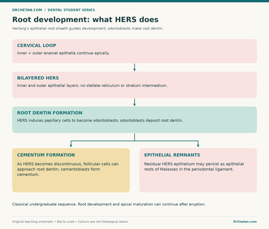
Figure 5. A process map of root development. It distinguishes the inducing epithelium from the cells that deposit root dentin and cementum.
Is root formation complete when a tooth erupts?
Not necessarily. A newly erupted permanent tooth can have an incompletely developed root and an open apex. Root development and apical maturation continue after clinical emergence.
Eruption is a coordinated developmental process involving the tooth’s surrounding tissues and bone remodelling. It should not be explained simply as “the root grows and pushes the tooth out.” For examinations, distinguish crown completion, eruption, root elongation, and apical completion as related but different landmarks.
Primary teeth, permanent successors, and permanent molars
Primary teeth arise at individual sites along the dental lamina. Permanent successor teeth develop from the successional lamina, an epithelial extension associated with the primary tooth germ on its lingual side.
The permanent incisors and canines replace their primary counterparts. Permanent premolars replace primary molars. Permanent molars do not replace primary molars: they are non-succedaneous teeth and develop from posterior extension of the dental lamina.
This is an important link between embryology and the tooth-identification guides in this series. A “primary premolar” is not part of the normal human primary dentition, and an adult first molar is not simply the replacement of the primary first molar.
Epithelial remnants: Serres versus Malassez
| Name | Main developmental association | Where they may remain |
|---|---|---|
| Epithelial rests of Serres | Remnants of the dental lamina | Gingival and jaw tissues |
| Epithelial rests of Malassez | Remnants associated with HERS | Periodontal ligament around the root |
The useful memory link is dental lamina ? Serres; root sheath ? Malassez. Their names do not make the structures interchangeable. In a developmental diagram, first establish whether you are discussing the original tooth-germ connection or the epithelial apparatus of root development.
How to recognise developmental stages in a slide
A histological section is a slice through a three-dimensional structure. The angle and level of the section affect the outline. Begin with tissue relationships, then identify the stage using several features.
| Stage | Features that favour it | What not to assume |
|---|---|---|
| Bud | Rounded epithelial outgrowth, epithelial connection, surrounding ectomesenchymal condensation | That the mature four-layer enamel organ is already obvious |
| Cap | Concave enamel organ over a papilla; surrounding follicle; emerging epithelial layers | That a shallow cup necessarily contains formed enamel |
| Early bell | Deeper concavity; four enamel-organ layers; increasingly specialised IEE and papillary cells | That every location already contains mineralised tissue |
| Advanced bell | Differentiated odontoblasts and ameloblasts with developing dentin/enamel in advanced crown regions | That all regions of the same crown are equally mature |
| Root-forming region | Bilayered HERS near the growing root; root dentin and developing supporting tissues | That HERS includes all four bell-stage layers |
For practice, the Histology Guide virtual slide linked in the references demonstrates tooth development in a fetal pig specimen. Its species is explicitly identified; it should not be described or reproduced as a human specimen. The diagrams in this article are original teaching schematics rather than micrographs.
Three transient names students encounter
- Enamel knot: a transient signalling centre involved in morphogenesis.
- Enamel cord: a strand of epithelial cells that may be described in relation to the knot and the enamel organ in early developmental sections.
- Enamel niche: an apparent arrangement produced by sectioning the curved dental lamina; do not treat it as a fourth principal component of the tooth germ.
Do not expect all three to be identifiable in every section. Their visibility and interpretation depend on the specimen and plane of section.
Why these stages matter clinically
Developmental biology helps distinguish changes in tooth number, shape, matrix quantity, mineral quality, and root pattern. However, a developmental defect cannot usually be assigned to one exact week or isolated cellular event from its appearance alone.
| Developmental concept | Example of the distinction it helps explain |
|---|---|
| Initiation and early patterning | Formation or absence of a tooth germ, and variation in tooth number |
| Crown morphogenesis | Altered crown outline or cusp pattern |
| Matrix secretion | Enamel hypoplasia involves reduced enamel quantity |
| Mineralisation and maturation | Enamel hypomineralisation concerns mineral quality rather than simply thickness |
| Root patterning | Variation in root length, form, or furcation pattern |
These are educational associations, not a diagnostic chart. Genetic effects, environmental influences, timing, duration, and the tissues involved can interact. The basic habit is to ask whether a problem concerns how much tissue formed, what form it took, or how it matured.
A simple revision method
First draw a cap-shaped enamel organ over a papilla and place a follicular outline around both. Name the origin and principal contribution of each component. Then expand one wall of the bell-stage organ and label OEE, SR, SI, and IEE in the correct order.
Next explain the hard-tissue sequence aloud: preameloblast–papilla interaction, odontoblast differentiation, predentin and first dentin, then enamel secretion. Finally, return to the cervical loop and explain how two epithelial layers become the root-guiding sheath.
This routine connects the diagrams instead of making you memorise five unrelated pictures. When a label is wrong, ask whether you confused cell origin, tissue position, developmental role, or chronology.
Practice questions
These are original revision questions for understanding and viva preparation, not reproduced past-paper questions.
| No. | Question |
|---|---|
| 1 | What do the names bud, cap, and bell primarily describe? |
| 2 | Which two tissue populations interact during early odontogenesis? |
| 3 | Name the three principal components of the tooth germ. |
| 4 | What is the difference between the dental and vestibular laminae? |
| 5 | What feature changes an epithelial bud into a cap-shaped enamel organ? |
| 6 | Which component gives rise to dentin and pulp? |
| 7 | Which enamel-organ layer gives rise to the ameloblast lineage? |
| 8 | List the four bell-stage enamel-organ layers from outside towards the papilla. |
| 9 | Where is the stratum intermedium located? |
| 10 | What is the difference between histodifferentiation and morphodifferentiation? |
| 11 | Which is formed first at the developing crown interface: dentin or enamel? |
| 12 | Does that sequence mean that preameloblast differentiation starts only after dentin forms? |
| 13 | What is predentin? |
| 14 | How does apposition differ from mineralisation? |
| 15 | What does the enamel knot principally do? |
| 16 | Which layers form HERS, and which two bell-stage layers are absent from it? |
| 17 | Does HERS directly secrete all the root dentin? |
| 18 | Which epithelial remnants are associated with dental lamina and with HERS? |
| 19 | Do permanent molars develop as successors of primary molars? |
| 20 | Does eruption prove that the root and apex have completed development? |
Answers with explanations
- Morphological changes in the developing enamel organ and tooth germ. They are not an alternative set of names for tissue secretion alone.
- Oral epithelium and neural crest-derived ectomesenchyme. Their reciprocal interactions guide development and differentiation.
- Enamel organ, dental papilla, and dental follicle/sac. They have different positions and contributions.
- Dental lamina participates in tooth formation; vestibular lamina contributes to the oral vestibule. Both are epithelial structures, but their outcomes differ.
- An indentation of the deep epithelial surface through differential growth. The enamel organ forms a cap over the papilla.
- Dental papilla. Peripheral cells become odontoblasts; central tissue develops into pulp.
- Inner enamel epithelium. Not every enamel-organ cell becomes an ameloblast.
- OEE ? stellate reticulum ? stratum intermedium ? IEE. Beyond the epithelial interface lies the papilla before hard-tissue deposition.
- Between the stellate reticulum and IEE. It is closely associated with enamel-forming cell function.
- Cell specialisation versus development of form. Odontoblast differentiation illustrates the first; crown and cusp patterning illustrates the second.
- Dentin. Initial dentin formation precedes adjacent enamel matrix secretion.
- No. Epithelial differentiation and signalling already participate in odontoblast induction; distinguish cell differentiation from matrix-deposition order.
- Unmineralised dentin matrix. It is secreted by odontoblasts before mineralisation produces dentin.
- Apposition adds matrix; mineralisation deposits mineral within it. Enamel maturation involves further changes in mineral, protein, and water.
- Acts as a transient signalling centre. It helps organise morphogenesis; it is not a finished enamel cusp.
- Inner and outer enamel epithelia form HERS; SR and SI are absent. It is a bilayered epithelial root-development structure.
- No. It guides and induces root development; odontoblasts produce root dentin.
- Serres with dental lamina; Malassez with HERS. Malassez rests are found in the periodontal ligament.
- No. Permanent molars arise from posterior dental-lamina extension; permanent premolars replace primary molars.
- No. Root elongation and apical maturation can continue after eruption.
Final revision checklist
Be able to explain the shape stages, the three tooth-germ components, the four enamel-organ layers, the first-dentin-before-first-enamel sequence, and the role of HERS. For each label, state where it is, what it comes from, and what it does.
The aim is to connect a changing shape with a changing cell population and a developing tissue. Once those relationships are clear, a tooth-development diagram becomes a story you can explain rather than a collection of names to memorise.

Leave a Reply