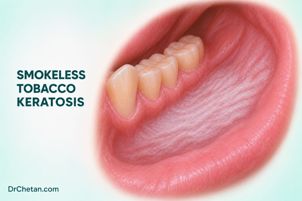
A patient who holds chewing tobacco in the lower buccal sulcus has a pale, wrinkled patch at the contact site. The exposure and location suggest a placement-associated reaction. A focal red area at one edge, however, raises a separate question about the tissue. Dental students should learn to document both the background change and the features that determine urgency, then check what happens after the visit.
Establish what touches the mucosa
Ask what the patient calls the product, what it contains and how it is used. Smokeless tobacco covers products with different processing methods, nicotine delivery and carcinogen content. In India, a patient may use tobacco with lime, a commercial mixture, or a betel quid with tobacco added separately. Ask about areca nut independently. An areca-containing product can be carcinogenic even when it contains no tobacco. Record frequency, years of use, duration in the mouth and usual placement. Where ingredients are uncertain, preserve that uncertainty in the record.
Ask whether the patient alternates sides, moves the product during the day or recently switched formulations. A lesion that corresponds closely to the habitual site supports a local response. The relationship still needs to be assessed alongside surface, firmness, change over time and other diagnoses. Examine the opposite side, the rest of the accessible mucosa and the neck as appropriate. A separate lesion can occur away from the reported placement site.
Describe the appearance before naming the lesion
| Observation | What to record | How it helps |
|---|---|---|
| Thin pale or grey area | Site, outline, relation to the sulcus and product placement | Establishes whether the distribution fits exposure. |
| Wrinkled or corrugated surface | Extent, thickness and whether it feels soft or firm | Helps compare the same area at reassessment. |
| A separate red, ulcerated or firm focus | Exact location, dimensions and progression | May require prompt assessment on its own merits. |
| Bilateral or distant changes | Sites on both sides and any other exposures | Broadens the differential beyond one placement site. |
An area of smokeless tobacco keratosis can have a relatively soft, pale or wrinkled appearance. More conspicuous keratin change can be seen with prolonged use. Frictional keratosis, leukoplakia, lichenoid disease and other white lesions can overlap in appearance. The label should follow assessment rather than replace it. If a lesion includes a focal red or firm area, describe the focal finding separately and decide whether it needs diagnostic assessment.
Interpret risk and regression in context
IARC’s prevention handbook includes smokeless tobacco keratosis among classified potentially malignant disorders with a qualification about differences among products. A study of one product and population cannot provide a single risk estimate for all placement-site lesions. Cancer risk relates to the exposure, while the diagnosis of an individual abnormality depends on examination and, when indicated, biopsy. Nicotine dependence, carcinogen content and the visible degree of keratosis also describe different features.
An often cited study reassessed young US military trainees after six weeks of enforced abstinence from smokeless tobacco. Most of the observed lesions resolved in that setting. The finding supports reassessment of selected lesions after exposure is addressed. It does not set a waiting period for a suspicious lesion or establish that the result applies to mixed areca and tobacco products used elsewhere. Study population, product and eligibility all matter.
Decide between prompt assessment and planned review

A persistent unexplained ulcer, induration, progressive growth or a concerning red or red-white area warrants prompt biopsy by an appropriately trained clinician or specialist referral. Record the reason for concern and the receiving service. If there are several components, use landmarks to show which one prompted the referral. A dated photograph with consent and scale can help, especially when the referral and biopsy are separated by time.
When findings are consistent with a low-risk local reaction and the clinician judges short observation appropriate, address the exposure and book a definite review. The ADA chairside guide advises biopsy or referral when a finding under watchful waiting persists beyond 10 to 14 days without a definitive diagnosis and treatment plan. That approach applies to selected observation. A suspicious focus warrants action at presentation. Track both the background change and any distinct focus.
If the biopsy report describes reactive keratosis, locate the specimen on the clinical map and check whether that finding explains the area of concern. A broad pale area can have a different histological appearance from a small red or indurated focus. If the clinical finding persists or progresses despite a reassuring result, discuss further assessment with the specialist.
Make the reassessment record usable
At the first visit, write where the lesion lies relative to fixed landmarks. A useful entry might describe diffuse keratosis in the left lower vestibule from the first premolar to the first molar, with a separate 5 mm red focus near the mesial border. State whether the patient can identify that area as a habitual site and which features were present before they changed products. At follow-up, record the same landmarks, dimensions and firmness. Photographs support the description when positioning and scale are reasonably consistent.
If the pale area recedes while an indurated focus remains, record those as different outcomes. The response of the surrounding mucosa supports part of the initial interpretation. The persistent focus still needs a diagnosis. Conversely, a patient who has been unable to stop using the product can still receive assessment and cessation support. Progress with quitting should never become a condition for examining a concerning lesion.
Connect the examination with cessation support
Offer tobacco cessation support and ask about areca use when relevant. A patient may stop one product and replace it with another. At follow-up, check each exposure and what happened at the lesion site. Record whether the person was able to access counselling or clinical cessation care. If a referral was issued for a lesion, verify its outcome separately.
A soft, diffuse change with a plausible contact site. The clinician documents a pale area corresponding to where the patient holds tobacco. There is no dominant firm or ulcerated focus. If the examination supports short review, discuss cessation and record the appointment. Reassess the same area and investigate persistence or change as indicated.
A new firm focus at the edge of a broad patch. The clinician documents both the diffuse change and the exact red, firm area. The focal finding is suspicious and prompts referral. A cessation conversation still belongs in the visit, while the diagnostic plan proceeds in parallel.

Leave a Reply