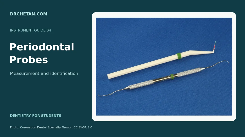
A periodontal probe is a graduated instrument used to assess periodontal tissues. Understanding its markings is essential: probes can look alike while using different graduation patterns.
Parts and general design
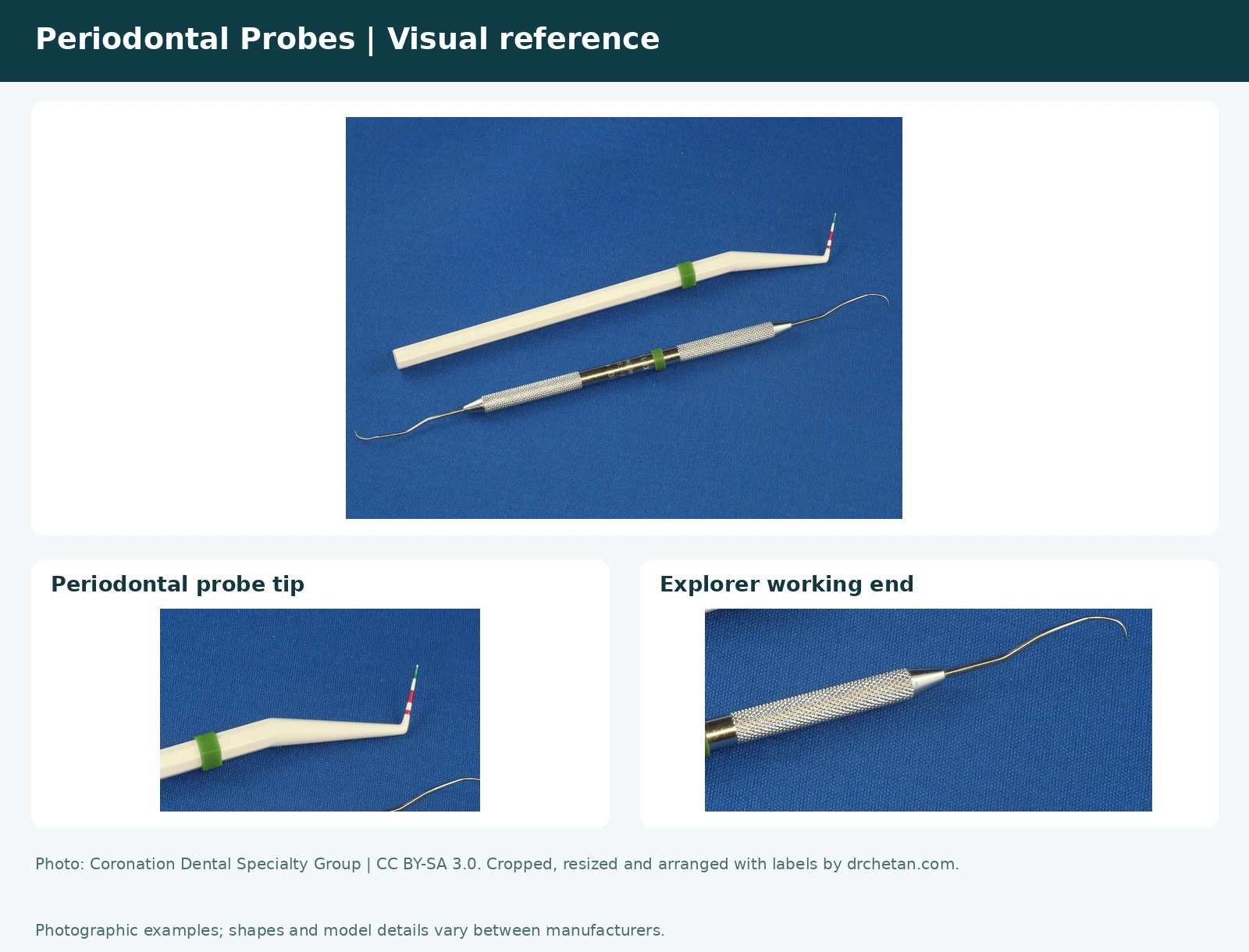
Identify the handle, shank and slender working end. A periodontal probe has a measuring tip and markings along the working end. The markings should be read according to the particular model, not guessed from a different instrument.
Photographic reference
UNC-15 probe
The UNC-15 is commonly graduated in millimetres to 15 mm. Coloured bands or markings help the operator recognise depths. Inspect the actual instrument and manufacturer’s guide because colour presentation can vary.
Williams probe
The traditional Williams pattern is marked at 1, 2, 3, 5, 7, 8, 9 and 10 mm, with gaps in the sequence. Learn to recognise its markings as a pattern rather than counting every visible space as one millimetre.
WHO/CPITN probe
The WHO/BPE probe has a 0.5 mm diameter ball tip and a black band from 3.5 to 5.5 mm. WHO clinical variants can include a second band from 8.5 to 11.5 mm. The ball tip and bands assist screening; check which version is in your kit.
Nabers furcation probe
The curved Nabers probe is designed to assess furcation involvement. Its curved working end and purpose distinguish it from a straight periodontal measuring probe. Some versions have markings; check the specific pattern.
Probe versus explorer
A periodontal probe has a measuring function. An explorer is designed for tactile assessment and typically has a fine pointed working end. A combined instrument can have one of each on opposite ends.
What does the measurement mean?
Probing depth is measured from the gingival margin to the base of the sulcus or pocket. Clinical attachment level uses a fixed reference, usually the cementoenamel junction (CEJ), to describe the position of attachment. The gingival margin can move with swelling or recession, so the two measurements are not interchangeable.
For example, a 4 mm probing depth with the gingival margin 2 mm apical to the CEJ gives 6 mm clinical attachment loss. When the margin lies coronal to the CEJ, the calculation must account for that position instead of automatically adding recession. This distinction is explained in SDCEP’s periodontal parameters guidance.
Screening versus detailed charting
A WHO probe is used in systems such as the Basic Periodontal Examination (BPE). BPE summarises findings to help decide the level of further assessment. It does not by itself diagnose periodontitis or replace site-specific periodontal charting. The original CPITN terminology and BPE describe examination systems; they are not names for every periodontal measurement.
Full assessment can include probing depths, gingival margin position, bleeding, furcations and mobility, interpreted with the history and other findings. A deep reading may be affected by gingival enlargement; it does not alone prove attachment loss. SDCEP also describes gentle probing and the limits of BPE.
Common marking patterns
- UNC-15: millimetre graduations from 1 to 15; bands commonly highlight 4–5, 9–10 and 14–15 mm.
- Williams: 1, 2, 3, 5, 7, 8, 9 and 10 mm. The missing 4 and 6 mm lines are intentional.
- WHO: ball tip and a 3.5–5.5 mm band; verify additional bands on the specific model.
- Nabers: curved furcation probe; marked versions commonly use 3, 6, 9 and 12 mm.
Semmelweis University’s instrument teaching material illustrates these probe types.
Viva questions
What is the UNC-15?
A periodontal probe commonly graduated to 15 mm.
What feature helps identify a WHO probe?
Its small ball tip and characteristic markings.
What is a Nabers probe used to assess?
Furcation involvement.
Why learn each probe’s markings?
Different patterns use different scales and bands, so readings depend on the actual instrument.
Does a deep pocket reading automatically mean periodontitis?
No. The gingival margin position and evidence of attachment loss must be assessed with the wider clinical findings.
Why should a probe be used gently?
Excess force can cause discomfort and alter tissue penetration. A light, controlled force and correct positioning improve the usefulness of the reading.
Further reading
Continue learning in the Dental Instruments and Dental Students collections.
References checked 31 October 2026. Educational identification guide for use alongside textbooks and supervised clinical teaching.
Featured-image photograph: Coronation Dental Specialty Group — source photograph (CC BY-SA 3.0). Resized and arranged with title; adapted visual: CC BY-SA 4.0.

Leave a Reply