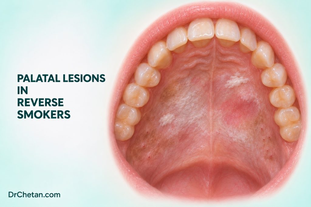
A patient says they smoke a chutta. The next question is how they hold it. In reverse smoking, the lit end is held inside the mouth, exposing the palate directly to heat and smoke. The resulting palatal findings are associated with a risk of oral cancer and appear in international classifications of oral potentially malignant disorders. A pale hard palate with red duct openings in a person who smokes conventionally can have a different clinical interpretation. The dental task is to identify the exposure, map the palatal findings and decide what needs prompt assessment.
Ask how the product is used
Ask which product is used and how it is held. A local product name such as chutta can refer to a tobacco roll, but the name alone cannot tell you whether the person places the burning end inside the mouth. Record current and past practice, frequency and years of use. Ask about other smoked and smokeless products, areca and alcohol separately. If the history is unclear, use neutral descriptive questions rather than assuming the practice from the person’s place of residence or age.
Older studies documented reverse smoking and palatal lesions in some rural communities in Andhra Pradesh. These reports remain useful for understanding the association and the clinical appearance. Their recruitment, place and period matter when interpreting any estimates. They do not establish how common reverse smoking or its associated lesions are in India today. Record the exposure history in the patient in front of you.
Distinguish background heat change from a suspicious focus
IARC’s oral neoplasia atlas illustrates a spectrum of palatal changes associated with reverse smoking, including diffuse keratosis and areas of altered colour or surface. The whole accessible palate should be examined, including the hard palate, the junction with the soft palate and any visible extension. Describe whether the finding is diffuse and symmetrical or focal and irregular. A dominant red patch, a persistent ulcer, a firm area or a growing mass needs separate assessment.
| Presentation | What can help distinguish it | What to document |
|---|---|---|
| Conventional smoker’s palate | Diffuse pale or grey keratotic hard palate with small red dots corresponding to inflamed salivary duct openings can fit nicotine stomatitis | Product and technique, distribution and any focal change that differs from the background. |
| Palatal changes in a reverse smoker | Direct exposure of the palate to the lit end, with a range of diffuse and focal findings | The exposure, exact distribution and suspicious red, white, ulcerated or firm areas. |
| A focal lesion on either background | Other inflammatory, infective, traumatic, potentially malignant or malignant causes remain possible | Size, colour, texture, firmness, duration, progression and the reason for concern. |
Nicotine stomatitis, also called smoker’s palate, is generally a heat-related response in conventional smoking. The red dots can be prominent minor salivary duct openings. The ordinary pattern often improves after the heat exposure stops. Palatal lesions associated with reverse smoking are classified separately as an oral potentially malignant disorder. Either patient can still have a suspicious focal lesion, and tobacco exposure remains important in both histories.
Examine and document the exact site
Use good lighting and inspect the hard and soft palates and the rest of the accessible oral mucosa. Note the side, relation to the midline, anterior or posterior location and any extent across the hard–soft palate junction. Describe the surface and palpate where appropriate, including for a submucosal mass or induration. Record whether a coating can be removed, whether an ulcer is present and whether a firm area extends beyond the visible change.
Use a dated photo with consent and scale when it helps, while retaining a written description. “Diffuse palatal whitening” and “10 × 7 mm firm red patch just left of the midline in the posterior hard palate” can both belong in the same chart. The second description gives a specialist a specific target for assessment. A small palatal swelling or ulcer can also reflect a salivary, dental or other process. Give the referral the differential as well as the exposure history.
Decide when a lesion needs prompt assessment
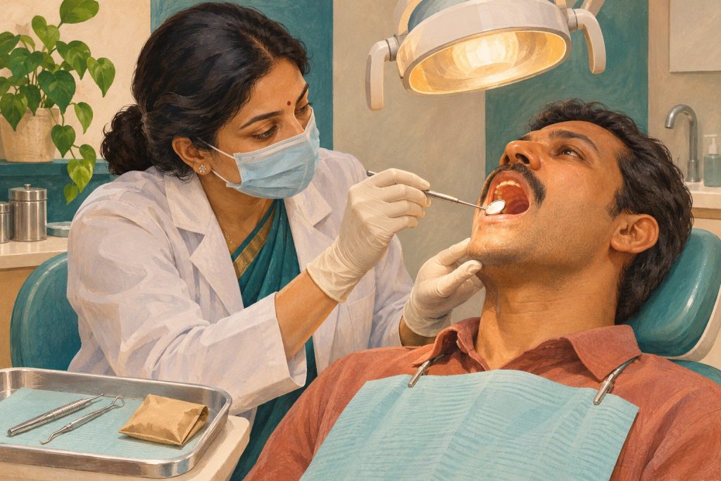
A persistent focal red or mixed plaque, ulcer, mass, induration or progressive change warrants prompt biopsy by a suitably trained clinician or referral when it appears suspicious. The presence of a smoking habit can make palatal findings more important to examine. It cannot establish the tissue diagnosis. When a focal change lies in a wide area of keratosis, identify the most concerning area on the referral and provide prior reports or photos when available.
If there is a convincing low-risk heat-related pattern and the clinician judges short observation appropriate, document the baseline, address the exposure and arrange reassessment. The ADA chairside guide advises biopsy or referral when a finding managed by watchful waiting persists beyond 10 to 14 days without a definitive diagnosis and treatment plan. Suspicious findings warrant prompt action at presentation. Cessation and diagnostic assessment can proceed together.
Offer cessation support and track the result
Explain the oral relevance of the smoking technique and the wider health risks of tobacco. Ask what makes stopping difficult and offer appropriate behavioural support or referral for tobacco cessation. For a patient who uses more than one product, ask about each separately at follow-up. A switch in product or technique changes the exposure history and should be documented.
Follow the lesion as well as the habit. If it was referred, confirm whether the patient reached the service and whether a biopsy result or other assessment is pending. If clinical observation was appropriate, compare the same site and document what resolved and what persists. New firmness, bleeding, ulceration or growth needs reassessment even when the diffuse whitening is improving.
Read historical evidence with attention to the setting
Cross-sectional studies can show that an exposure and a lesion occur together in a population. They cannot establish the time course for every individual or provide a current national estimate. Studies from particular communities also reflect which products and techniques were used at the time. Use the historical evidence to recognise the practice and its association with palatal disease, and use the current examination to decide what needs assessment.
A concise clinical record can separate the exposure and the findings. “Reports smoking with the lit end inside the mouth” describes the technique. “Diffuse posterior hard-palate keratosis with a firm focal ulcer near the midline” describes what was observed. Add measurements, duration if known and the referral plan. The record makes clear why the focal lesion warrants attention within a wider background change.
If the clinical finding is close to the hard–soft palate junction, record the apparent centre and whether the posterior edge was visible. When the view is limited, say which part could not be assessed. The receiving team can correlate the exact location with the tissue result and imaging when indicated.
Apply the distinction to two cases
Diffuse palatal change in a conventional smoker. Examination shows diffuse pallor with several red duct openings and no dominant ulcer or mass. Clarify the smoking technique, map the finding and discuss cessation. If the overall findings support planned review, set a date and assess whether the palate changes after the exposure is addressed.
A focal ulcer in a reverse smoker. A patient describes holding the lit end of a tobacco roll inside the mouth. There is diffuse palatal keratosis and a new firm ulcer in one region. Document the exposure and both findings. Arrange prompt specialist assessment of the ulcer, and give the receiving clinician the exact site and reason for urgency.

Leave a Reply