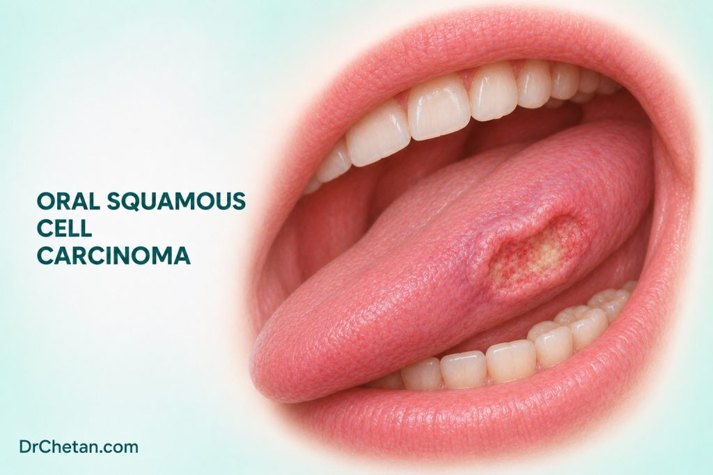
A patient returns after a lower molar extraction because the socket has enlarged and bleeds when touched. The surrounding gingiva is firm, and the change extends onto the lingual mucosa. A dental explanation may still be possible, but the progression calls for an assessment that can establish a tissue diagnosis. This is one way oral squamous cell carcinoma, or OSCC, can first appear in dental practice. The student’s task is to recognise the concerning pattern, document the site and organise prompt assessment. Biopsy, imaging and staging then answer different questions.
Define OSCC by its tissue and site
OSCC is an invasive malignant epithelial tumour of the oral cavity with squamous differentiation. The oral cavity includes the mobile tongue, floor of mouth, gingiva and alveolar ridges, inner cheek, hard palate, retromolar trigone and mucosal lip. A squamous carcinoma arising in the tongue base or palatine tonsil is oropharyngeal. Its diagnostic testing and staging may follow a different pathway. When a lesion crosses a boundary, describe the likely epicentre and involved structures rather than assigning the whole lesion to whichever surface is easiest to see.
Conventional OSCC is the focus here. The WHO oral cavity and mobile tongue chapter also lists verrucous carcinoma and carcinoma cuniculatum as distinct entities. They can require careful clinicopathologic correlation, particularly when a superficial specimen does not capture the interface with underlying tissue. Other squamous carcinoma forms show different microscopic patterns, such as spindle cell or basaloid morphology. Surface roughness or a warty appearance can help describe a lesion. Histological type is established by pathology.
Tobacco, areca nut and alcohol are important exposures. A history of an oral potentially malignant disorder can alert the clinician to a site that has changed or to risk elsewhere in the mouth. OSCC can also present without a known precursor or recorded exposure. Use the history to inform the examination and prevention conversation, and judge the current finding on its own clinical features.

Recognise the presentation and its mimics
The pattern varies by site and extent. A lateral tongue tumour may appear as a persistent ulcer with an indurated margin. An early lesion can be painless. A gingival lesion can resemble periodontal disease, a reactive growth or a socket that has failed to heal. A floor-of-mouth lesion may become clearer when the tongue is elevated and the area is palpated. Restricted tongue movement, trismus, persistent bleeding, difficulty swallowing and a cervical lump may accompany more extensive disease or a primary that is hard to inspect.
| Presentation | Alternative explanations to consider | Details that change the assessment |
|---|---|---|
| Persistent tongue ulcer | Trauma, inflammatory ulceration or infection | Growth, induration, fixation and whether a plausible irritant explains the whole finding. |
| Red-white patch with a firm area | An OPMD with or without dysplasia, inflammatory change or invasive disease | Site of the firm focus, change since prior examination and relation to earlier biopsies. |
| Enlarging gingival or socket lesion | Reactive tissue, infection, a jaw lesion or another malignancy | The chronology, tooth and bone findings, extension beyond the socket and whether the clinical course fits the dental explanation. |
| Exophytic or warty growth | Reactive or benign epithelial proliferation and verrucous tumours | Base, depth, surrounding mucosa and which areas may need sampling. |
| Cervical lump with little visible oral change | Inflammatory node, a concealed head and neck primary or other disease | Duration, site, mobility, throat symptoms and limits of the examination. |
Record dimensions, exact subsite, side, relationship to the midline and any deep fixation, mucosal extension or functional change. Examine the rest of the oral cavity and accessible neck. The referral should capture relevant risks, prior biopsies and the reason for concern. A suspicious lesion calls for prompt biopsy by a suitably trained clinician or referral to a service that can establish the diagnosis. Treatment of a possible dental cause should be part of a documented assessment, especially when the course or examination is atypical.
Connect the biopsy to the clinical question
A biopsy asks whether the lesion is invasive and what type of tumour or alternative process is present. The approach depends on location, size, suspected diagnosis and operator expertise. In a heterogeneous lesion, the site and depth of sampling matter. Representative tissue should let the pathologist assess the epithelial and stromal interface. A superficial fragment or necrotic material may leave the question of invasion unresolved. Provide the specimen site and the clinical appearance, and state what the assessment needs to resolve.
When the report and clinical findings disagree, look at the sample and the lesion together. A superficial biopsy reported as reactive may be accurate for the tissue submitted even though the underlying lesion remains suspicious. Reassess the site and sampling, communicate with pathology and arrange repeat or specialist-directed biopsy as indicated. A photograph or site map can help establish which part of a broad lesion the specimen represented.
On microscopy, invasive OSCC can form irregular nests, cords or individual tumour cells in the stroma. Keratinisation varies. Well-differentiated examples may show keratin pearls and intercellular bridges, while poorly differentiated areas can show less obvious squamous maturation. Invasion is the central distinction from epithelial dysplasia. The pathologist interprets the architecture with the cytology, specimen quality and clinical context. Histologic differentiation is one feature of the tumour and does not supply its anatomic stage.
Read the resection report beyond the diagnosis line
An initial incisional biopsy may establish the diagnosis without showing the deepest point of invasion or the full lesion. A resection report can provide a more complete account. ICCR’s oral cavity dataset covers resections and excisional biopsies within its scope. Review the primary tumour report alongside the neck specimen report where applicable.
| Report item | What it adds |
|---|---|
| Primary site and tumour dimensions | Establishes the anatomic context and contributes to primary tumour classification. |
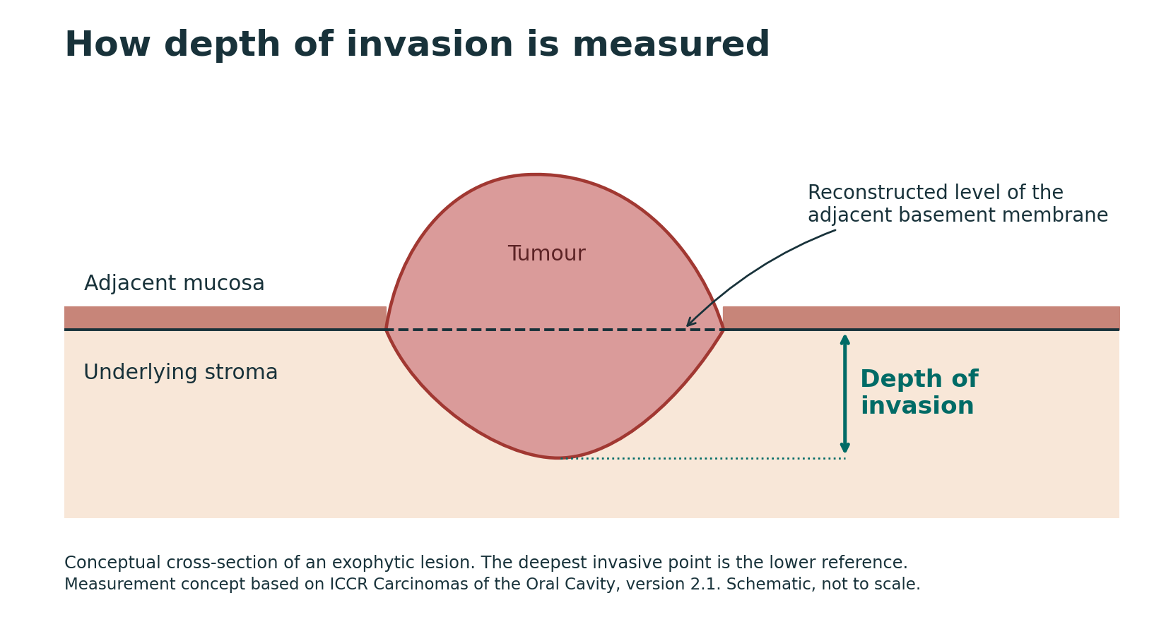
| Depth of invasion | Measures the deepest invasive point from a reconstructed level of adjacent normal mucosal basement membrane. It contributes to oral cavity SCC staging. |
| Margins | Records the relation of invasive carcinoma and relevant epithelial abnormalities to the surgical edges. Review the named margin, the distance where reported and the specimen context. |
| Perineural or lymphovascular invasion | Describes tumour involving nerves or vascular spaces. These findings contribute to postoperative risk assessment. |
| Nodal findings and extranodal extension | Describes involved nodes and whether tumour extends beyond a nodal capsule. Details contribute to stage and adjuvant treatment decisions. |
Depth of invasion and tumour thickness use different reference points. An exophytic tumour can project above the adjacent mucosal level, making its thickness greater than its measured depth of invasion. An ulcerated lesion can lose surface tissue and make thickness measured from the ulcer base smaller than the depth referenced to the expected mucosal level. In both cases, the pathology report should document the measurement within the protocol used.
Put site and pathology into the staging workup
Clinical staging combines examination and appropriate investigations before definitive treatment. CT or MRI may clarify deep extent, relation to bone and the neck. Specialist teams choose the imaging according to the clinical question and suspected disease extent. Needle sampling can help assess a suspicious node. Pathologic staging, when available, adds findings from resection and nodal assessment. A small visible surface lesion can have appreciable depth, and a clinically node-negative neck can contain occult metastases.
The AJCC 2026 table retains the eighth edition for oral cavity cancers. The primary tumour category incorporates tumour size, depth of invasion and, when present, qualifying local extension. The stage group combines the primary tumour, regional nodal and distant metastasis categories. For a teaching example, an oral cavity SCC measuring 18 mm with a pathologic depth of invasion of 7 mm and no qualifying invasion of adjacent structures fits the size and depth criteria for pT2 in the eighth edition. The stage group still requires nodal and distant information.
Read the staging example as a sequence of evidence
Consider the 18 mm lesion first at a dental examination and then after surgery. The dental record might describe an ulcer on the right lateral mobile tongue, a firm base and a neck without palpable nodes. The clinical team can refine the extent with imaging and directed assessment. Only the resection measurement in this example supplies the 7 mm pathologic depth of invasion. If a subsequently examined node contains metastatic SCC, the nodal classification must reflect the pathological findings even if there was no suspicious node on the initial dental examination. The same patient can therefore have two legitimate records of extent, each tied to what was known at a particular time.
Pay attention to which specimens the pathology report represents. A report covering the primary resection can describe site, margins and depth. A separate neck report can describe nodal levels, numbers of involved and examined nodes, and extranodal extension where present. Bringing both reports to the multidisciplinary discussion avoids inferring a final stage from the primary tumour measurement alone.
Understand treatment decisions without prescribing from stage alone
Resectable oral cavity SCC is commonly managed with surgery when appropriate. The team plans tumour clearance, function and reconstruction, and considers how to assess or treat the neck. Treatment choice also depends on primary site, extent, patient fitness, expected effects on speech and swallowing, and patient preferences. Definitive radiotherapy or other approaches may be appropriate in selected circumstances.
The neck needs separate consideration even when clinical examination and imaging show no suspicious nodes. An Indian randomised trial in selected early, clinically node-negative oral cancers compared elective neck dissection at primary surgery with therapeutic dissection after nodal relapse. It demonstrated better outcomes with the elective approach in that study population. ASCO guidance supports elective neck dissection in defined oral cavity scenarios. Specialist centres may use sentinel node biopsy for selected early cases where expertise and a suitable pathway exist. The trial and guidelines inform multidisciplinary decisions for the particular patient.
After surgery, the multidisciplinary team reviews features such as pathologic stage, margins, nodal involvement, extranodal extension, perineural invasion and lymphovascular invasion. Postoperative radiotherapy may be indicated for adverse features. Positive margins and extranodal extension are high-risk findings that can lead to consideration of postoperative concurrent chemoradiotherapy in suitable patients. The precise recommendation depends on the pathology, whether additional surgery is feasible, expected benefit and patient fitness.
Follow the handoffs that affect dental care
Before treatment, coordinate with the oncology team on dental infection control, caries and periodontal assessment, and the timing of any invasive procedures. Anticipate the functional effect of resection or reconstruction. Where head and neck radiotherapy is planned, the team can advise which structures will receive dose and how dental treatment should be timed. After cancer treatment, monitor and support oral hygiene, nutrition, mouth opening, salivary symptoms and dental rehabilitation within the coordinated care plan. A new progressive mucosal lesion or neck finding during follow-up warrants reassessment.
Return to the opening example. Record the site as the left or right mandibular alveolus and note where the change reaches the adjacent mucosa. Describe how quickly the socket enlarged, the character of the tissue, induration and neck findings. Arrange prompt assessment and establish who will track the referral and pathology result. If the biopsy confirms OSCC, the diagnosis resolves the tissue question. The next team still needs the anatomical extent, neck and distant assessment, and a treatment plan that includes function and dental care.

Leave a Reply