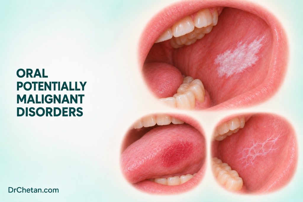
A dentist sees a white patch on the lateral tongue at a routine visit. The patient has no pain and cannot say when it appeared. The first task is to describe the finding and consider what might explain it. If the patch remains unexplained, the dentist must decide whether assessment can safely begin with a short review or requires prompt biopsy or referral. The term oral potentially malignant disorder, or OPMD, helps describe cancer risk across a group of disorders. It cannot establish the tissue diagnosis of the patch in front of you.
Separate the clinical label from the tissue diagnosis
An OPMD is a disorder associated with an increased risk of developing oral cancer. Some present as a single patch. Others affect several sites or involve widespread mucosal changes. Risk varies across and within diagnoses, and carcinoma can arise in the previously recognised lesion or elsewhere in the mouth. The clinical label conveys a risk context and helps plan assessment and follow-up. It does not specify what is present microscopically.
Oral epithelial dysplasia describes microscopic architectural and cellular abnormalities in the epithelium. The pathologist assigns a grade within the reporting framework used. Dysplasia can be present in a clinically defined OPMD, and an OPMD may be diagnosed even when the sampled tissue lacks dysplasia. Invasive squamous cell carcinoma involves growth into underlying tissue. A lesion referred as suspected leukoplakia or erythroplakia can already contain invasive disease when first sampled. An initial descriptive diagnosis is therefore provisional until the clinical findings and any biopsy have been reconciled.
An oral lesion can be clinically suspicious even without a classic OPMD pattern. An unexplained indurated ulcer, a progressive mucosal mass or a suspicious cervical node deserves appropriate assessment regardless of whether an OPMD label fits. The action depends on the overall findings, including the extent, history, limitations of the examination and any evidence of progression.
Classify the presentation before narrowing the diagnosis
| Presentation | Diagnoses to consider | Findings that inform assessment |
|---|---|---|
| Persistent white plaque | Leukoplakia after other causes are excluded, frictional keratosis or another white lesion | Wipeability, site, relation to trauma, uniformity, texture and change over time. |
| Predominantly red or mixed red and white lesion | Erythroplakia, erythroleukoplakia or an inflammatory or infective mimic | Margin, persistence, focal induration, ulceration and the part of the lesion that looks most abnormal. |
| Progressive white plaques at several sites | Proliferative verrucous leukoplakia, lichenoid or reactive mimics | Map the sites, record surface evolution and compare prior biopsy locations and results. |
| Blanching and restricted mouth opening | Oral submucous fibrosis in an appropriate clinical context | Areca products and duration, palpable bands, measured opening and any focal change. |
| Bilateral reticular change or focal erosions | Oral lichen planus, oral lichenoid lesions or another immune-mediated disorder | Symmetry, distribution, symptoms, prior treatments and any new focal induration or persistent ulceration. |
This pattern-based approach guides examination and referral. The international consensus and IARC handbook also discuss actinic cheilitis, palatal lesions in reverse smokers, oral lupus, oral manifestations of chronic graft-versus-host disease and rare inherited conditions. IARC includes smokeless tobacco keratosis with a qualification about variation by product. A patient can have more than one process at the same time, such as diffuse fibrosis with a new focal ulcer. Assess the focal change on its own merits.

Recognise the major disorders
Leukoplakia and its common mimics
Oral leukoplakia is a clinical diagnosis of exclusion for a predominantly white plaque of uncertain risk after other recognised disorders that explain the appearance have been excluded. Document whether the plaque is relatively uniform or has mixed colour, nodularity or verrucous change. A cheek lesion at the occlusal line with a convincing mechanical source has a different starting differential from a persistent irregular plaque on the ventrolateral tongue. Assess both the relation to trauma and what happens after the source is addressed when a short review is clinically appropriate.
Risk is higher in some presentations. A systematic review associated nonhomogeneous appearance, greater size, lateral tongue location and epithelial dysplasia with a higher likelihood of malignant transformation across included studies. The studies used different follow-up durations and patient selection criteria. Their pooled estimates describe groups. A benign-looking surface, a low-risk location or a reassuring past result can inform the assessment, but current clinical change still deserves attention.
A systematic review and meta-analysis of 55 studies reported a pooled malignant transformation proportion of 6.64% for oral leukoplakia, with a 95% confidence interval of 5.21% to 8.21%. Between-study heterogeneity was high. The number represents the pooled experience of the included cohorts, with varying eligibility criteria and periods of observation. Use lesion characteristics, histology and change over time to assess an individual case. Two patients carrying the same clinical label may need different assessment because the lesion’s site, surface, extent, histology and trajectory are different.
For a concrete comparison, a homogeneous plaque with no dysplasia on representative sampling may be followed under a defined specialist plan. A large irregular plaque with a new red or firm focus may require additional tissue assessment even if another part of it was benign on biopsy. Document the observation that changes the plan instead of relying on a numerical threshold drawn from a pooled study.
Erythroplakia and mixed lesions
Erythroplakia is an unexplained red mucosal patch after other definable conditions have been excluded. A mixed red-white patch may be described as erythroleukoplakia when the clinical criteria are met. Inflammatory, traumatic, vascular and infective findings can also appear red. The diagnostic task is to reconcile the colour with the lesion’s margin, consistency, site and history, then take appropriate action when it remains suspicious. Erythroplakia studies include cases with severe dysplasia or carcinoma already present on initial biopsy. That finding answers a different question from the risk that a non-invasive lesion will progress during follow-up.
Proliferative verrucous leukoplakia
Proliferative verrucous leukoplakia is recognised through its clinical course and clinicopathologic correlation. Early lesions can look relatively flat and white. Over time, more sites may become involved and some areas become thicker or verrucous. The clinician should preserve a dated map and record which part of which lesion was sampled. A single pathology report can describe only the submitted tissue at that time. Serial examinations and biopsies can show change that is invisible in an isolated report. Progressive multifocal disease merits specialist assessment and a follow-up plan that accounts for the whole mouth.
Oral submucous fibrosis
Oral submucous fibrosis is associated strongly with areca nut use. In a patient reporting burning with spicy food and progressive difficulty opening the mouth, look for mucosal blanching and palpable fibrous bands where examination permits. Measure interincisal opening and note which surfaces could not be examined. Ask about supari, pan masala, betel quid and other local preparations by name and record whether tobacco is also used. Restricted access makes the search for a new focal lesion more difficult. A persistent ulcer or a firm focal change within the fibrotic mucosa requires independent assessment.
Oral lichen planus and lichenoid disorders
Bilateral reticular change can support a diagnosis of oral lichen planus in an appropriate clinical context. Erosions, symptoms, asymmetric disease and focal changes require correlation with the history and examination. Record the background pattern separately from any persistent ulcer, induration or new red-white area. Reviews report differing estimates of malignant transformation depending on which diagnostic criteria were used and how dysplasia and lichenoid lesions were handled. This makes accurate classification and follow-up more informative than quoting a single number to an individual patient.
Choose between prompt assessment and short review
When a lesion is suspicious at the first visit, arrange prompt biopsy by a suitably trained clinician or referral to a specialist who can obtain it. Features that heighten concern include an unexplained firm or indurated ulcer, a persistent red or red-white patch, progressive growth, fixation, spontaneous bleeding and a concerning cervical node. Their significance depends on context, and the absence of one feature does not cancel the others. Record how quickly the patient should be seen, where the referral was sent and how completion will be checked.
A brief reassessment can be appropriate when there is a convincing benign explanation and no concerning clinical feature. For example, remove a clear source of mechanical injury, document the baseline and book a definite review. The ADA’s 2026 chairside guidance advises biopsy or referral if an abnormality under watchful waiting persists beyond 10 to 14 days without a definitive diagnosis and treatment plan. Continued abnormality, progression or new concern requires escalation according to the clinical findings. NICE’s separate UK pathway includes unexplained oral ulceration lasting more than three weeks among its referral criteria. In a suspicious presentation, clinical findings can justify prompt assessment at the first visit.
Biopsy selection matters in a large or heterogeneous lesion. Communicate the concerning site, clinical differential and relevant prior biopsy results. A specialist may sample more than one area where the appearance varies, or arrange repeat assessment if a benign or low-grade result does not account for the clinical findings. Light-based adjuncts and vital stains do not replace tissue assessment. The ADA limits the role of cytology to defined contingency circumstances, and any result must be followed through within the recommended pathway.
Follow the patient after the report returns
Start by comparing the tissue diagnosis with what was seen. “No dysplasia identified” describes the sampled tissue. In a persistent heterogeneous lesion, reassess whether the specimen represented the area that raised concern. If dysplasia is present, management takes account of the grade, site, extent, clinical appearance, procedure morbidity and ability to return for care. Selected lesions may be observed under a documented surveillance plan. Others merit further sampling or intervention. Even after treatment, the clinician should continue to assess the treated site and the surrounding and contralateral mucosa.
Agree a surveillance interval with the treating team, state who will examine the patient and give specific reasons to return earlier. Useful examples include growth, a new red area, ulceration, increasing firmness, new restriction in mouth opening and a neck lump. At each visit, compare with the prior description, map or consented photographs. Continue to discuss exposure cessation and arrange support where needed. A follow-up plan should remain usable when a patient moves between a primary dental clinic and a specialist service.
Work through two chairside decisions
A new red focus within a long-standing white lesion. A patient presents with a persistent lateral tongue plaque and brings a previous report that found no dysplasia. Today there is a firm red area that was absent from the earlier photograph. Record the new focus, review where the previous tissue was taken and arrange prompt specialist reassessment or appropriate biopsy. The new focus and prior sample represent different observations. The referral needs both.
Limited opening with a new unilateral ulcer. A patient with diffuse blanching and a history of chewing areca nut develops a painful unilateral ulcer that cannot be fully seen. Record the measured opening, the suspected location and the examination limits. Arrange prompt assessment of the ulcer and address the exposure and fibrosis in the wider care plan. Difficulty seeing the lesion adds a reason to establish how specialist assessment will be completed.

Leave a Reply