
A patient points to the back of the tongue and says that swallowing hurts. A dental mirror shows a small ulcer on the posterolateral mobile tongue. Are the symptom and the visible finding related? And if the ulcer proves malignant, which cancer pathway applies? The answer starts with identifying where the lesion arose. Mobile-tongue squamous cell carcinoma belongs to the oral cavity. A squamous carcinoma arising at the tongue base belongs to the oropharynx. Two lesions a short distance apart can therefore have different pathology testing, staging and treatment workups.
Map the oral cavity from front to back
Start at the mucosal lip and labial vestibule, then inspect both buccal sulci, buccal mucosa, gingiva and alveolar ridges. Inspect the hard palate and note where it gives way to the mobile soft palate. At the lower molars, look behind the last tooth to examine the retromolar trigone. The part of the tongue that can be moved forward for examination includes the tip, dorsum, lateral borders and ventral surface of the mobile tongue. The sulcus terminalis marks the anatomical division between the mobile tongue and tongue base. Circumvallate papillae lie immediately anterior to it, and their appearance alone should not be used to locate the origin of a lesion that spans the boundary.
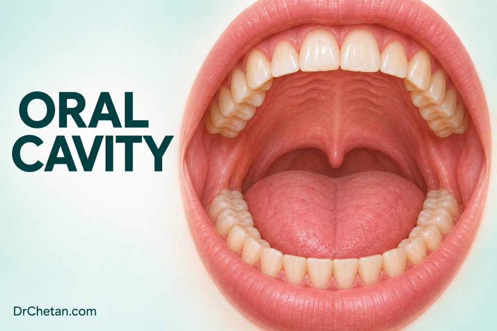
Record both the oral cavity subsite and the surface involved. “Left lateral mobile tongue” is more useful than “tongue.” “Left ventrolateral mobile tongue extending onto the floor of mouth” adds a second observation about extent. Two adjacent structures can move differently as the patient protrudes or elevates the tongue. Observe how the visible lesion relates to those movements, then palpate where appropriate to assess induration and whether the finding appears fixed to deeper tissues. Examination can suggest the epicentre. Imaging and pathology may change the final assignment.
| Oral cavity site | What to record | Why it matters |
|---|---|---|
| Mucosal lip | Upper or lower lip, surface of origin and extension toward dry vermilion or skin | Lip lesions can fall under different reporting rules depending on where they start. |
| Buccal mucosa and vestibule | Side, level relative to the teeth and extension into a sulcus or commissure | A buccal primary can reach the adjacent alveolus. Record the apparent point of origin. |
| Gingiva and alveolar ridge | Upper or lower, adjacent teeth or extraction socket, surface and fixation | A mucosal lesion can extend into jawbone. Tooth mobility or a socket finding alone does not establish bone invasion. |
| Mobile tongue | Lateral, ventral, dorsal or tip, distance from midline and mobility | The report needs to distinguish mobile tongue from tongue base and describe any midline or floor-of-mouth extension. |
| Floor of mouth | Side, midline, relation to frenulum, duct region and mandible | Inspection alone can underestimate a deep or submucosal finding. |
| Hard palate | Side and relation to the midline, alveolus and hard–soft palate junction | A palatal lump has a differential beyond surface squamous malignancy, including minor salivary tumours. |
| Retromolar trigone | Relationship to last mandibular molar, ascending ramus, buccal mucosa and tonsillar region | A lesion can extend posteriorly, so record what you can see and where examination stops. |
These fields help a specialist correlate the referral with a biopsy label and imaging. The distinctions between mucosal lip, dry vermilion and skin also make it easier for the pathologist to apply the correct protocol.
Identify the oropharyngeal landmarks
The oropharynx lies behind the oral cavity. It includes the tongue base, palatine tonsils and tonsillar region, soft palate and oropharyngeal walls. The anterior tonsillar pillar is the mucosal fold over the palatoglossus muscle. The posterior pillar overlies the palatopharyngeus, and the palatine tonsil lies between them. At the tongue base, lingual tonsillar tissue can add an irregular contour. A visible asymmetry deserves correlation with symptoms and palpation findings where appropriate. The visible part of the throat provides only a partial examination of the region.
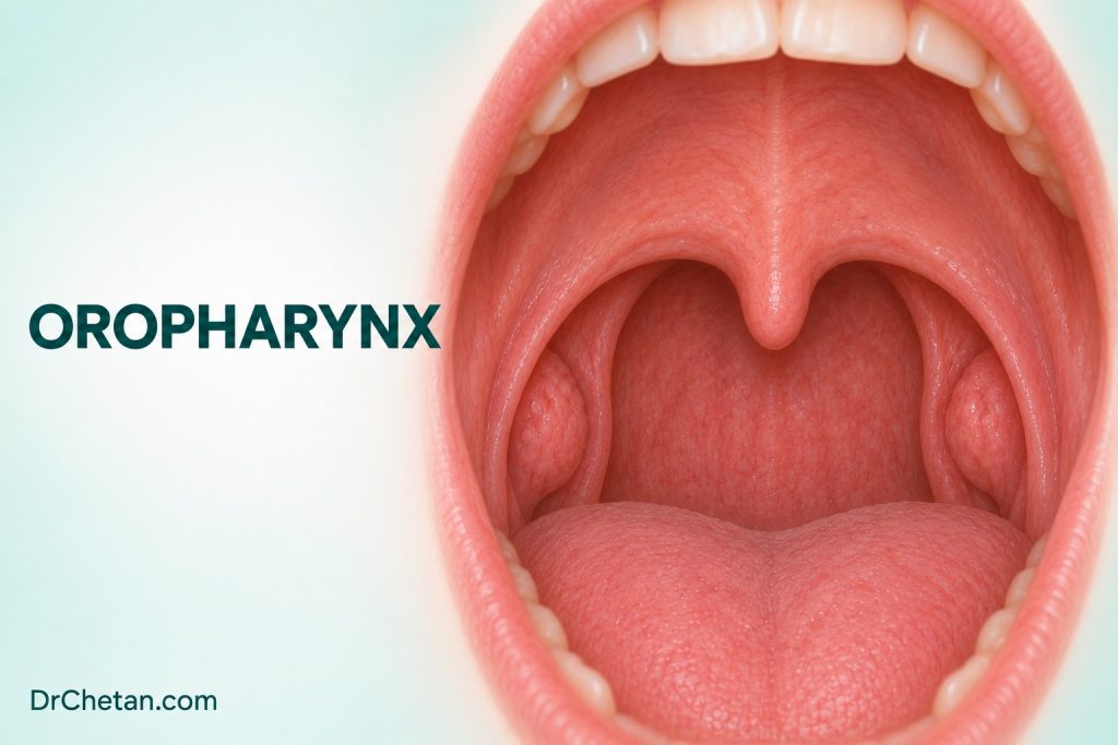
Behind the mandibular molars, the retromolar trigone can sit close to the anterior tonsillar pillar. The hard–soft palate junction is another border that looks different according to mouth opening and viewing angle. When a lesion spans a junction, describe the centre of the visible or palpable abnormality, which structures are crossed and how much confidence you have in your impression. If the posterior edge disappears from view, say so. Labeling the whole lesion “oral” because it is visible through the mouth can direct the receiving team toward the wrong assumptions.
Symptoms can point beyond the area visible to a dentist. A persistent unilateral throat symptom, pain on swallowing or a neck mass can accompany an oropharyngeal primary, including one that is small or concealed. An ordinary chairside examination does not visualise the entire tongue base or the interior of a tonsillar crypt. Explain the examination limit to the patient and arrange specialist assessment when symptoms or examination findings warrant it. An ENT or head and neck team can decide whether flexible endoscopy, imaging or tissue assessment is needed.
Examine and document what you can see
Use a systematic visual and tactile examination of the mouth and accessible neck. Good light, a mirror and a gauze hold on the anterior tongue can improve visualisation. Ask the patient to move the tongue side to side and elevate the tip. Inspect both lateral borders and the ventral tongue. Compare the right and left floor of mouth and palpate where appropriate. Look at the soft palate and visible tonsillar region without forcing the examination when gagging, pain or trismus limits access. Document the limits of examination and the features actually observed.
For a mucosal lesion, record the greatest visible dimension, colour and surface, margin characteristics, bleeding and induration. Include the relationship to the midline and which landmarks have been crossed. If an image can be obtained with consent, use a consistent view and scale, identify the side and date, and keep it within the clinical record. Write the site in words even when a photograph is available. A close crop may show surface detail without conveying which part of the tongue or palate it came from.
If you detect a cervical lump, describe its side, location, size and mobility. Add the time course and any relevant oral or throat symptoms to the referral. Palpable normality and radiological normality also describe different assessments. A neck that feels normal can still harbour occult nodal disease after cancer is diagnosed. When an adult presents with a persistent neck mass of uncertain origin, specialist workup may involve directed examination, imaging and needle sampling.
Write a referral that another clinician can reproduce
A useful site description can read: “Left posterolateral mobile tongue, approximately 14 × 9 mm on the visible surface. Ulcer with a firm anterior margin on gentle palpation. It is separate from the visible floor of mouth and does not reach the midline. The posterior edge cannot be fully seen because of limited opening. Symptoms began four weeks ago and have progressed.” These observations give the receiving clinician a starting location, the most concerning finding and an explicit examination limit. They do not claim a tissue diagnosis or define how far the lesion extends under the surface.
When a site is uncertain, draw the visible extent on a simple oral chart and write a suspected site of origin in words. For multiple findings, number them and describe each separately. Label any tissue sample with the same site description so that the referral, photographs and pathology can be reconciled. A label such as “posterior tongue lesion 2” becomes useful only when the chart says which surface, which side and how the lesion relates to the mobile portion and the visible tonsillar region. If a node is the first specimen, document that the mucosal primary is uncertain and record the site of nodal sampling separately.
Why site changes HPV testing and staging
High-risk HPV has an established role in a subset of oropharyngeal squamous cell carcinomas, particularly those arising in tonsillar tissue of the palatine tonsil and tongue base. CAP recommends high-risk HPV assessment for newly diagnosed oropharyngeal SCC within the guideline’s scope. p16 immunohistochemistry serves as a surrogate in appropriate tissue settings. CAP specifies circumstances for HPV-specific testing, including situations where morphology and p16 findings are discordant and particular geographic or specimen contexts. The pathology team applies those details to the case in front of it.
For a typical SCC with a confirmed oral cavity origin, CAP advises against routine high-risk HPV testing for prognostic purposes. The context matters when the primary site is unclear, especially if the first tissue available is a cervical node. For example, a positive p16 stain in a nodal metastasis may help direct the workup. CAP recommends high-risk HPV testing when metastatic SCC of unknown primary is found in a cervical node. For non-cytology tissue specimens, CAP advises HPV-specific testing or p16 immunohistochemistry followed by HPV-specific testing when p16 is positive. A dental referral should give the actual site observations so that the oncology team can direct that workup.
The AJCC 2026 current staging table retains the eighth edition oral cavity chapter. AJCC Version 9 applies to HPV-associated oropharyngeal carcinoma from 1 January 2026. The previous p16-positive oropharynx terminology may still appear in older records or teaching materials. Document the tumour site, tumour type, HPV context where applicable and the staging version used by the treating team. In oral cavity SCC, depth of invasion contributes to the primary tumour category. The oropharyngeal system has its own site- and HPV-context-specific rules. A measurement or category taken from an oral cavity example cannot be carried straight into an oropharyngeal case.
| Localisation question | What the distinction changes |
|---|---|
| Mobile tongue or tongue base | The cancer site assignment, HPV workup for SCC and the appropriate staging chapter. |
| Hard palate or soft palate | Whether the lesion belongs anatomically to the oral cavity or oropharynx. The tissue diagnosis still determines the reporting framework. |
| Retromolar trigone or tonsillar pillar | The proposed primary site and which extent needs further examination when it cannot be seen at chairside. |
| Oral primary visible or cervical node without visible primary | How the specialist investigates an occult primary and selects an appropriate specimen for testing. |
Apply the map to three referrals
A mobile-tongue ulcer approaching the posterior border. An indurated ulcer is visible on the left lateral tongue when the patient protrudes it. Its posterior margin becomes difficult to see on retraction. Document the visible lesion on the lateral mobile tongue and the uncertain posterior extent. Prompt biopsy by an appropriately trained clinician or specialist referral is indicated when the findings are suspicious. A symptom described as “back of tongue pain” does not override what you can localise on examination.
A neck lump with a visible tonsillar asymmetry. An adult reports a persistent unilateral neck lump and discomfort on swallowing. The oral cavity examination is unremarkable, but the left tonsillar region looks asymmetric. Describe the asymmetry, neck finding and duration. Arrange prompt specialist assessment. The receiving team can evaluate the upper aerodigestive tract and select imaging and needle sampling as appropriate. The absence of a visible oral cavity ulcer is part of the record. It does not resolve the origin of the neck finding.
A palatal lesion at the junction. A firm swelling crosses from posterior hard palate to soft palate. Record the visible centre, surface changes, symmetry and boundaries, and distinguish the firm submucosal component from any overlying ulcer. The workup needs both a site assignment and a tissue diagnosis. A minor salivary carcinoma and a surface squamous carcinoma can share a general location yet require different reporting and staging considerations. If inspection cannot establish the epicentre, preserve that uncertainty in the referral.
A concise referral can use this structure in a single paragraph: location and side, duration and change, lesion measurements and concerning features, adjacent structures and limits of view, neck findings, and the reason for referral. When working across the oral cavity and oropharynx, the most useful early information is a reproducible description of what is visible, what is palpable and which boundary remains uncertain.

Leave a Reply