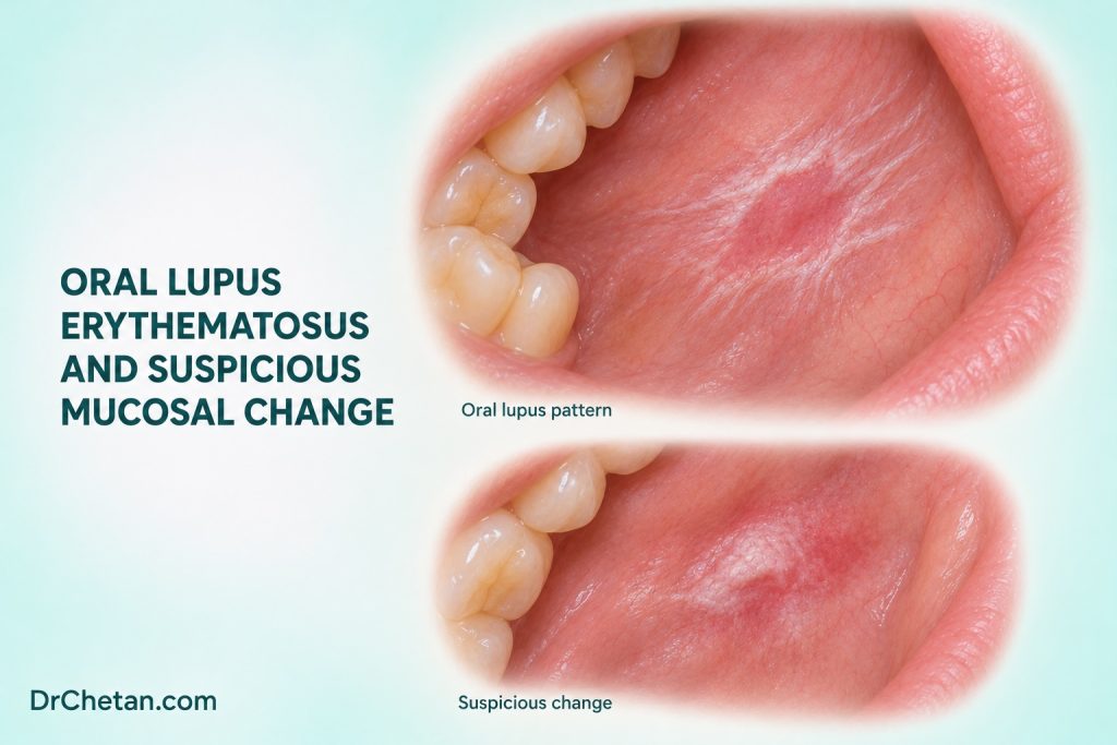
A patient with lupus develops a red patch on the hard palate. A second patient has a chronic lip lesion with a scaly border and a new firm focus. Both histories deserve attention, but the diagnosis of lupus cannot establish what caused the current finding. At chairside, document the distribution and the focal change, consider several explanations and coordinate assessment with the relevant team.
Start with the clinical context and distribution
Lupus erythematosus can involve oral mucosa in systemic or cutaneous disease. Presentations include ulcers, erythema and lesions with white keratotic or striated borders. Some discoid lesions can show a central area of erythema or atrophy with peripheral white striae. The palate, buccal mucosa and lips are among the sites to examine. Morphology and distribution vary, and an apparent lupus lesion may overlap with oral lichen planus, a lichenoid reaction or another inflammatory or neoplastic process.
Ask about the type of lupus if known, previous oral findings, current systemic treatment and recent changes. Record oral symptoms and whether they correspond to systemic disease activity or an independent clinical event. Consider trauma, infection and medication effects. The treating team can help clarify immunosuppression, relevant laboratory findings and procedural safety when tissue assessment is being planned. Do not assume that oral symptoms establish a systemic flare.
| Finding | Possible interpretations | What to record or clarify |
|---|---|---|
| Red or ulcerated palatal area | Lupus-associated change, trauma, infection or another focal process | Duration, exact site, symptoms and whether the appearance is progressing. |
| Red plaque with peripheral white striae | Discoid or lichenoid disease, among other possibilities | Distribution, symmetry, other affected sites and the clinical history. |
| Chronic scaly lip change | Lupus-related cheilitis, actinic change or another cause | Apparent surface of origin, sun exposure and any focal firmness or crusting. |
| New focal induration, persistent ulcer or growth | A suspicious focus that can coexist with inflammatory disease | Dimensions, consistency, relevant neck findings and the referral plan. |
Use biopsy to resolve the right question
A biopsy may help distinguish lupus-associated inflammation from other interface disorders, infection, epithelial dysplasia or invasion. Oral lupus can show interface changes that overlap with lichenoid disease. Histological features should be interpreted with the clinical distribution and medical history. If direct immunofluorescence is appropriate, the specialist can arrange a suitable specimen and handling. Communicate whether the question concerns a representative background lesion or a focal area suspicious for malignancy.
When the report arrives, identify which part of the lesion was sampled. A result describing inflammation in one site cannot resolve a new firm focus at another. Compare the visible and palpable findings with prior records. If the clinical finding and pathology disagree, arrange further assessment through the treating team. A familiar autoimmune diagnosis should remain part of the context rather than becoming the default explanation for every lesion.
Explain what the cancer evidence measures

International consensus includes oral lupus erythematosus among oral potentially malignant disorders. An observational review found an association between systemic lupus and oral cancer at the patient-population level and reported prevalence estimates in studies of discoid lupus. These are different endpoints. Neither establishes how often a biopsy-confirmed oral lupus lesion will progress, or predicts what will happen at an individual site. Study design, selection, disease definitions and treatment context affect interpretation.
The practical implication is to reassess suspicious change. A longstanding lesion can change, and a new process can arise within or next to an established background disorder. Record whether there is persistent ulceration, progressive thickening, firmness, unexplained bleeding or a change in colour or extent. A relevant exposure history can inform risk assessment, while the clinical finding governs whether prompt biopsy or referral is needed.
Write the differential so the next clinician can act
A useful referral might describe a persistent 9 mm ulcer on the right hard palate with a firm margin, note when it appeared and document how it differs from previously recorded mucosal changes. List relevant lupus treatment and immunosuppression, then state what the referral should resolve. If several areas are present, number them or use clear landmarks so the biopsy request can identify the same focus. A note that simply says “oral lupus” leaves the receiving clinician without a description of the feature that changed the decision.
A typical inflammatory pattern and a new suspicious finding can be assessed on separate timelines. Review ongoing symptoms with the medical team and arrange prompt assessment of the focal lesion as indicated. If a previously recorded ulcer improves, document the site and extent of resolution, including any residual firmness or surface change. This gives future clinicians a baseline and keeps a partial response from being mistaken for resolution of the whole concern.
Coordinate follow-up and keep the focal finding visible
Explain what the examination established and which question remains open. Coordinate with oral medicine and the patient’s lupus team when necessary. A patient may have a plan for systemic symptoms and a separate plan for the oral lesion. Document who will check the referral or pathology result and when a definite reassessment is due. For a suspicious lesion, arrange prompt assessment rather than waiting to see whether systemic treatment improves the mouth.
A stable bilateral inflammatory pattern. The patient reports intermittent sensitivity and has no dominant firm or ulcerated site. Record the clinical distribution and compare with earlier records. Review medication, supportive care and follow-up with the treating team. Explain which focal changes should bring the patient back sooner.
A new firm focus within a chronic lip lesion. Record the exact surface, side, size and relationship to the background change. Arrange prompt specialist assessment if the focus is suspicious. Send the relevant history and earlier pathology, and track whether the patient reached the receiving service.

Leave a Reply