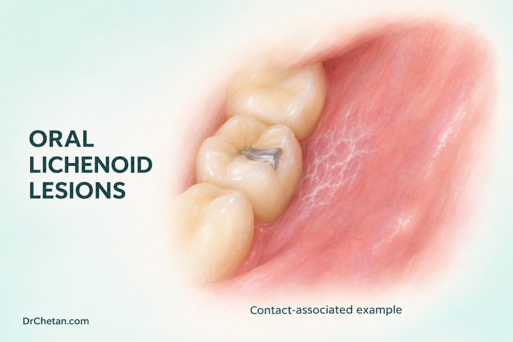
A patient has a white striated patch next to a restoration on one cheek. Another has similar changes on both sides and recently started a new medicine. In both cases the pattern is lichenoid, but the contact site and timing are clues rather than proof of a cause. The dental record should describe what the clinician sees, which exposures are plausible and what evidence would change the working diagnosis.
Use the term as a working diagnosis
Oral lichenoid lesions can resemble oral lichen planus clinically and histologically. The term is used in different ways in published criteria. It can include a lesion with a possible contact or drug trigger and cases that resemble OLP but fail to meet a particular set of diagnostic criteria. State the suspected explanation and degree of certainty. If a trigger is unclear, record the clinical and histological observations that support a lichenoid pattern.
| Pattern or context | What it suggests | What to check next |
|---|---|---|
| Localised lesion in direct contact with a restoration | A contact-associated reaction is possible | Map the contact and examine areas that do not touch the material. |
| Onset after a change in medication | A drug-related reaction belongs in the differential | Confirm the chronology, other exposures and indication for the medicine with the treating clinician. |
| Multifocal bilateral striae | Oral lichen planus is a consideration | Compare both sides, ask about other mucosal symptoms and look for discordant focal changes. |
| Lichenoid change after allogeneic transplantation | Oral chronic graft-versus-host disease may be relevant | Ask about the transplant and coordinate assessment with the treating team. |
| Persistent focal ulcer, red plaque, induration or growth | Another process may coexist with lichenoid inflammation | Arrange prompt assessment or biopsy when the lesion is suspicious. |
Examine the whole accessible mouth rather than only the area that prompted the visit. Record symmetry, sites, dimensions, surface and firmness. Ask about dental work, products applied to the mouth, prescription and over-the-counter medicines, symptom onset and previous pathology. A complete timeline helps distinguish a long-standing lesion that was first noticed after treatment from one that plausibly began after exposure.
Investigate contact without assuming material allergy
A lesion that directly touches a restoration is more suggestive of a local contact process than a broadly symmetrical pattern. Confirm how the cheek or tongue meets the restoration at rest or during movement. Check for a mechanical explanation, and record whether similar lesions occur elsewhere. The presence of a restoration by itself is common and cannot establish causation.
Specialists sometimes use patch testing to investigate suspected contact allergy. Results need interpretation against the distribution, material and clinical context. A positive skin patch test indicates sensitisation under the conditions of that test. Its relevance to the oral lesion still needs assessment. Some selected patients improve after replacement of a directly contacting restoration, but the evidence includes observational studies with heterogeneous selection and outcomes. Consider restoration condition, the risks of replacement and the plan for reassessment before proposing dental treatment.
If a material change is agreed, document the appearance before treatment and how the lesion changes afterwards. Improvement can support a contact explanation. Persistent or progressive change may warrant reconsideration or further assessment. A suspicious focus deserves diagnostic attention before relying on a material change to explain it.
Investigate a possible medicine reaction safely
Build the medication timeline with start dates, dose changes and approximate lesion onset. Ask about earlier episodes and alternative triggers. Published evidence for individual drug causes is uneven and often comes from case reports. Some reactions may appear after a delay. Temporal association can support a hypothesis, but other causes should still be assessed.
Coordinate with the prescriber when a medicine is a plausible contributor. The prescriber can assess the indication, alternatives and consequences of changing treatment. The patient should have a clear plan for observation and reassessment if the medication changes. A persistent red or ulcerated focus with concerning features should be assessed in parallel. Do not wait for a possible drug effect to resolve before addressing a suspicious finding.
Use biopsy to answer a specific question

Lichenoid interface inflammation can occur in more than one disorder. When the clinical diagnosis is uncertain or a focal area is suspicious, communicate the distribution, exposures, clinical differential and exact sample location to the pathologist. A biopsy can identify dysplasia, invasion or another diagnosis that changes the plan. Clinicopathological correlation is especially important when the report says “lichenoid mucositis” and the examination shows a focal progressive lesion.
If the main presentation is desquamative gingivitis or blistering, consider other immune-mediated disorders. An oral medicine specialist can decide whether direct immunofluorescence or other investigations are appropriate. The location and handling for these samples may differ from routine histology. State the clinical question rather than assuming that one type of specimen addresses all possibilities.
Document how you would test the working explanation
Set a defined review point before a suspected trigger changes. Record the exposure, lesion location, baseline symptoms and the clinical feature that should improve if the explanation is correct. A structured description can later show that the soreness improved while a plaque persisted, or that change occurred only at the contact site. These are more informative than describing the whole mouth as better.
Consider other causes when the course is unexpected. A persistent focal white plaque, candidal changes, lupus-related lesions or a blistering disorder can overlap with a lichenoid appearance. The clinician may need to revisit the clinical differential, the specimen or the need for specialist assessment. The decision depends on the remaining findings and the reliability of the original explanation.
Plan follow-up around the actual finding
Oral lichenoid lesions are included among oral potentially malignant disorders in international consensus. Transformation estimates vary with case definitions, whether epithelial dysplasia was present and length of follow-up. A patient with a documented contact-associated lesion and a patient with an unexplained dysplastic lichenoid lesion have different management questions. Explain what the biopsy and clinical course establish and what remains under assessment.
Record whether the suspected trigger was changed, the date of reassessment and which features should bring the patient back sooner. Use earlier descriptions or consented photographs to compare the same site. A persistent ulcer, firmness or progressive red or white change warrants reassessment even if other areas have improved. Check that the referral or biopsy result reaches the clinician who will act on it.
Where a plausible contact or medication association remains unconfirmed, it is reasonable for the chart to retain a working description such as “unilateral lichenoid lesion, possible contact association.” Include the planned next observation or investigation. If a specialist establishes an alternative diagnosis, update the record and communicate which follow-up is still needed.
Two decisions to practise
Unilateral change at a restoration. A patient has striae where a buccal surface contacts a restoration. Map the contact and examine the rest of the mouth. Discuss whether specialist assessment and a planned material change are appropriate. Record which findings would trigger reassessment earlier, especially a new firm or ulcerated focus.
New medication and a firm ulcer. A patient associates oral soreness with a newly started medicine, and examination shows a firm focal ulcer. Take the medicine history and coordinate with the prescriber. Arrange prompt specialist assessment of the ulcer. Both workups can proceed at the same time, with separate documentation of medication decisions and the lesion outcome.

Leave a Reply