A practical dental anatomy guide for BDS students: recognise incisors, canines, premolars, and molars; distinguish maxillary from mandibular teeth; and assess right and left using several anatomical clues.
You are handed an unlabelled tooth in a dental anatomy practical. You recognise that it is a posterior tooth, but is it a first premolar or a second premolar? Is it maxillary or mandibular? And how should you decide whether it belongs on the right or left?
The answer is not to search for one memorable feature and stop. Identify a tooth by a combination of features, examined from several surfaces. The overall crown, cusp arrangement, grooves, root form, and mesial–distal differences should tell a consistent story.
This guide gives you a repeatable method, followed by the distinguishing features of the permanent tooth types. It describes typical teaching morphology; real teeth vary. Wear, caries, restorations, fractures, and developmental variation can obscure the textbook pattern.
In this guide:
- What counts as a complete identification?
- Understand the terms before comparing teeth
- A five-step method for an unlabelled permanent tooth
- Incisors: broad upper crowns versus slender lower crowns
- Canines: compare crown bulk and lingual anatomy
- Premolars: compare cusps, grooves, and mesial features
- Molars: inspect the occlusal table before counting roots alone
- How to assess right and left without reversing the tooth
- A rapid reference to the 16 permanent tooth types
- Common confusions and how to resolve them
- How to present an answer in a practical or viva
- A useful practice routine
- Practice questions
- Final revision checklist
What counts as a complete identification?
A complete tooth name establishes the dentition, arch, patient’s side, and exact tooth type. For example, “permanent maxillary right first premolar” is more informative than “upper premolar.” Its FDI code is 14.
In a practical examination, also explain why you reached that answer. A useful response is: “I identify this as a permanent maxillary first premolar because of the two-cusp crown, mesial marginal groove, and mesial crown–root depression. I would use those mesial features to help establish the side.”
For an isolated specimen, you may be confident about the tooth type but less confident about the side. That is better than claiming certainty from a worn cusp or an unreliable root bend. In the mouth, use the tooth’s position and surrounding dentition as well as morphology.
Understand the terms before comparing teeth
| Term | Meaning in tooth identification |
|---|---|
| Maxillary / mandibular | Belonging to the upper / lower jaw |
| Mesial / distal | Towards / away from the midline of the dental arch |
| Facial | The outward-facing surface; labial for anterior teeth and buccal for posterior teeth |
| Lingual / palatal | Towards the tongue / towards the palate; palatal is used for the inner surface of maxillary teeth |
| Incisal / occlusal | Cutting edge of an anterior tooth / chewing surface of a posterior tooth |
| Cervical line / CEJ | The cementoenamel junction separating the anatomical crown and root |
| Cusp | A raised part of the crown with a cusp tip and surrounding slopes or ridges |
| Cingulum | A convexity in the cervical part of an anterior tooth’s lingual surface |
| Fossa / groove | A depression / a linear surface marking; identify its location and relationships |
| Mesiodistal / buccolingual | Width between mesial and distal surfaces / between buccal and lingual surfaces |
When you describe a tooth as “wide,” specify the direction. A crown can be narrow mesiodistally but relatively broad labiolingually. Also distinguish a root from a root canal: a single root can contain more than one canal. This article concerns external identification, not prediction of internal canal anatomy.
A five-step method for an unlabelled permanent tooth
Step 1: Orient the specimen
Locate the crown, cervical line, root or roots, and incisal or occlusal surface. Then identify the facial and lingual surfaces. Anterior lingual surfaces have structures such as a cingulum, marginal ridges, and a fossa; facial surfaces have a different contour.
Rotate the specimen through facial, lingual, mesial, distal, and incisal or occlusal views. A feature that is hard to understand from one angle may become obvious from another. Do not treat the first view as the whole tooth.
Step 2: Establish the tooth class
- Incisor: a cutting edge rather than a single pointed cusp or broad multicusped occlusal table.
- Canine: one principal cusp and a substantial single-rooted form in the usual specimen.
- Premolar: a smaller posterior occlusal table, usually with a buccal and lingual cusp arrangement; some have three cusps.
- Molar: a broad chewing table with several major cusps and commonly multiple roots.
These are starting patterns. For example, a mandibular first premolar’s small lingual cusp can make its buccal view look canine-like. Looking from the occlusal surface helps reveal its posterior-tooth architecture.
Step 3: Decide maxillary or mandibular
Use the clues specific to that tooth class. Maxillary incisors generally have a broader crown and more marked lingual anatomy than mandibular incisors. Maxillary molars typically have three roots and an oblique ridge; mandibular first and second molars typically have two roots and a different cusp–groove arrangement.
Premolars require a more specific comparison. The tiny lingual cusp of a typical mandibular first premolar, for example, is more helpful than a general statement about upper versus lower tooth size.
Step 4: Identify the member within the class
Ask whether the incisor is central or lateral, whether the premolar is first or second, or whether the molar is first, second, or third. Use comparisons rather than isolated lists: what makes this specimen more consistent with one member than its closest alternative?
Step 5: Assess the side and then assign the notation
Identify mesial and distal using the most reliable features preserved on that tooth. Orient it in the appropriate arch before deciding the patient’s right or left. Once you have the full name, assign the code using a named system.
Morphology establishes identity; notation records it. If you need a refresher on FDI, Palmer, and Universal notation, use the companion tooth-numbering guide in this series. Do not use an unexplained number as a substitute for identifying the specimen.
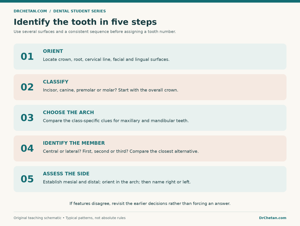
Figure 1. Work from broad classification to specific identity. Recheck earlier decisions if the later features do not fit.
Incisors: broad upper crowns versus slender lower crowns
There are eight permanent incisors: a central and lateral on each side of both arches. Their cutting edges distinguish them from the cusped canine form, but tooth-to-tooth differences are easiest to understand by comparison.
Maxillary central incisor
Look for a relatively broad mesiodistal crown with a substantial incisal edge. In a typical unworn labial view, the mesioincisal corner is sharper, while the distoincisal corner is more rounded. The distal crown outline is usually more convex than the mesial outline.
The lingual surface has a cingulum, marginal ridges, and a lingual fossa. The usual root is single and conical. An unworn newly erupted crown may show mamelons, but their presence or absence is not a dependable way to identify a central incisor by itself. [1,2]
Most useful comparison: versus the maxillary lateral incisor, the central generally looks broader and less rounded in crown form. Compare several features, because the lateral may have a relatively long root despite its smaller crown.
Maxillary lateral incisor
The crown is generally narrower and more rounded than that of the maxillary central. Its lingual fossa and marginal ridges can be more pronounced, and a lingual pit or palatogingival groove may be present.
The lateral incisor is variable: a peg-shaped form is a familiar example of altered crown morphology. Do not make a pit, groove, or a particular root bend mandatory for identification. Use the smaller rounded crown and its other surviving anatomical features together. [3]
Mandibular central incisor
This is a small, narrow-mesiodistal incisor with a comparatively symmetrical crown. The lingual surface is generally smoother and less strongly sculpted than that of a typical maxillary incisor.
Its incisal corners and mesial–distal crown contours can look very similar. That symmetry is useful for identifying the tooth type, but it makes deciding right versus left more difficult. The root is commonly flattened mesiodistally, with proximal longitudinal depressions.
Practical lesson: do not force a confident side assignment when a worn or unusually symmetrical specimen lacks clear evidence. Record the observations that support the central-incisor identification, then assess the side separately. [4]
Mandibular lateral incisor
The mandibular lateral is usually slightly larger and less symmetrical than the mandibular central. Its incisal edge commonly shows a distolingual twist: the distal part turns lingually, following the curve of the arch.
You appreciate this feature from the incisal view, not by looking only at the facial surface. A more rounded distal corner and asymmetry of the crown can support the identification. Again, wear can reduce the value of these clues. [4]
Incisor comparison for revision
| Tooth | Features to look for | Main confusion |
|---|---|---|
| Maxillary central | Broad crown; sharper mesioincisal and rounder distoincisal corner | Maxillary lateral |
| Maxillary lateral | Smaller, more rounded crown; often more marked lingual anatomy; variable form | Maxillary central |
| Mandibular central | Narrow crown; relatively bilateral symmetry; subdued lingual anatomy | Mandibular lateral |
| Mandibular lateral | Slightly larger, more asymmetrical crown; distolingual incisal twist | Mandibular central |
Canines: compare crown bulk and lingual anatomy
A typical permanent canine has a single principal cusp, a labial ridge, a lingual cingulum, and a long root. The mesial cusp ridge is generally shorter than the distal cusp ridge in an unworn permanent canine. This helps identify mesial and distal, but it is not a rule to apply blindly after cusp wear.
Maxillary canine
The maxillary canine is usually bulkier labiolingually and has more prominent crown relief than the mandibular canine. Its lingual ridge can divide the lingual surface into mesial and distal fossae, with a relatively marked cingulum and marginal ridges.
Its long, robust root supports the overall identification. However, do not decide “maxillary canine” solely because a specimen has the longest root among a handful of teeth on a tray: root completeness and individual variation affect that comparison. [5]
Mandibular canine
The mandibular canine usually appears narrower mesiodistally, with a comparatively long-looking, slender crown. The lingual anatomy tends to be less pronounced. From the facial view, the mesial crown outline often continues relatively straight into the mesial root outline.
The usual root is single, but variants, including divided roots, occur. Root form should support the crown-based identification, not overrule it simply because a specimen differs from a teaching model. [5]
| Feature | Typical maxillary canine | Typical mandibular canine |
|---|---|---|
| Crown impression | Bulkier and more strongly sculpted | Narrower and relatively slender |
| Lingual anatomy | More prominent ridge, fossae, and cingulum | Less pronounced relief |
| Mesial facial outline | More obvious crown–root contour change | Often a straighter crown–root continuation |
| Best approach | Combine lingual and facial views with root form | Combine slender form, smoother lingual view, and mesial outline |
Premolars: compare cusps, grooves, and mesial features
The permanent dentition has eight premolars. There are no primary premolars. “Bicuspid” is a familiar term, but it is not a reliable promise that every premolar has exactly two cusps: a mandibular second premolar may have three.
Maxillary first premolar
This tooth has a particularly useful set of identification features:
- A buccal and a lingual cusp, with the buccal cusp typically longer.
- A relatively angular or hexagonal occlusal outline.
- A mesial marginal developmental groove crossing the mesial marginal ridge.
- A mesial developmental depression that involves the crown and can continue onto the root.
- An unworn buccal cusp whose mesial cusp ridge is generally longer than the distal cusp ridge.
That last feature is a useful contrast with the usual permanent canine pattern. The maxillary first premolar’s buccal cusp tip is displaced distally rather than being simply centred between equal cusp ridges.
Two roots, buccal and palatal, are common, but single-rooted and other configurations occur. A one-rooted specimen is not automatically a second premolar. The crown and mesial features remain important. [6]
Maxillary second premolar
The maxillary second premolar generally has a more rounded occlusal outline. Its buccal and lingual cusps are more similar in height, the central developmental groove is shorter, and supplementary grooves can make the occlusal surface look more wrinkled.
It usually has a single root. The first premolar’s distinctive mesial marginal groove and marked mesial crown–root depression are not normally part of the same characteristic feature package here.
The practical question is not merely “one root or two?” Compare cusp-height difference, outline, groove pattern, and mesial surface. [6,7]
Mandibular first premolar
Think of a tooth with a dominant buccal cusp and a much smaller lingual cusp. The occlusal table slopes strongly towards the lingual side, and a pronounced transverse ridge is a useful feature in a typical specimen.
A mesiolingual developmental groove is a particularly helpful clue. The mesial marginal ridge slopes cervically towards the lingual side. From the buccal view alone, this tooth can resemble a canine; the small lingual cusp and occlusal architecture reveal its premolar identity. [8]
Avoid confusing “mesial marginal groove” and “mesiolingual groove” as though they are interchangeable names. Locate the feature on the specimen and describe what structure it crosses or separates.
Mandibular second premolar
Compared with the first, the mandibular second premolar has a better-developed lingual part of the crown and a more substantial occlusal table. It commonly has either:
- Three cusps: one buccal, one mesiolingual, and one distolingual; the major groove arrangement is Y-shaped.
- Two cusps: one buccal and one lingual; the groove pattern is commonly described as H-shaped or U-shaped.
In the three-cusp form, the mesiolingual cusp is usually larger than the distolingual cusp. The single buccal cusp and the distribution of the lingual cusps matter more than simply counting three cusps and assuming that the tooth must be a molar. [9]
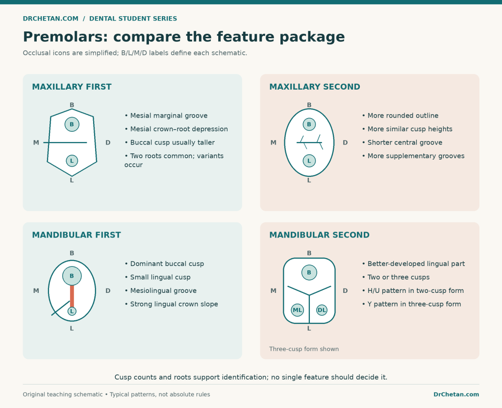
Figure 2. Compare a feature package: cusp proportions, mesial landmarks, occlusal outline, and the usual root pattern. These are typical patterns, not absolute rules.
Premolar comparison for revision
| Tooth | Most useful feature combination | A mistake to avoid |
|---|---|---|
| Maxillary first | Mesial marginal groove plus mesial crown–root depression; taller buccal cusp | Rejecting it because only one root is present |
| Maxillary second | More rounded outline; more similar cusp heights; shorter central groove and supplementary grooves | Assuming every one-rooted premolar is this tooth |
| Mandibular first | Dominant buccal cusp; small lingual cusp; mesiolingual groove; lingually sloping table | Calling it a canine from the buccal view alone |
| Mandibular second | Better-developed lingual portion; two- or three-cusp form; H/U or Y groove pattern | Assuming all specimens have three cusps |
Molars: inspect the occlusal table before counting roots alone
The permanent dentition can have 12 molars when all four third molars are present. Molar identification benefits from combining the occlusal outline, cusp arrangement, ridges, and roots. Do not count worn cusp remnants without looking for their relationships.
Maxillary first molar
The typical crown has four major cusps: mesiobuccal, distobuccal, mesiolingual, and distolingual. An oblique ridge connects the mesiolingual and distobuccal cusps, a key maxillary-molar landmark.
The occlusal outline is generally rhomboidal. The usual three roots are mesiobuccal, distobuccal, and palatal. The palatal root is the substantial lingual root, rather than a third buccal root.
A Carabelli trait may occur on the palatal surface of the mesiolingual cusp. Its expression varies from a small surface feature to a more conspicuous accessory cusp. Do not require a large Carabelli cusp in every first molar, or count it as one of the four principal cusps. [10]
Maxillary second molar
The second molar is commonly smaller, with greater reduction of the distal part of the crown, particularly the distolingual cusp. Some specimens have a four-cusp rhomboidal form; a reduced or absent distolingual cusp can produce a heart-shaped, three-cusp outline.
The roots are often less divergent and may lie closer together than those of the first molar. Their degree of separation is supportive evidence, not a guarantee. Also, a heart-shaped crown does not automatically identify a third molar. [11]
Maxillary third molar
Third molars are especially variable in crown size, cusp form, grooves, and roots. A reduced distolingual region, heart-shaped crown, and short or fused roots may support a maxillary third-molar interpretation, but none is compulsory.
Compare the whole specimen with first- and second-molar patterns. In a clinical setting, its position and imaging provide information that an isolated tooth on a practical tray cannot supply. [11]
Mandibular first molar
The usual mandibular first molar has five major cusps: mesiobuccal, distobuccal, distal, mesiolingual, and distolingual. From the buccal aspect, the distal cusp contributes to the familiar three-cusp buccal profile; from the lingual aspect, two lingual cusps are seen.
Its crown is generally wider mesiodistally than buccolingually. The usual two roots are mesial and distal, rather than buccal and palatal. Two buccal developmental grooves are associated with the three-part buccal cusp profile in the typical five-cusp form. [12]
Mandibular second molar
The typical mandibular second molar has four major cusps with a more regular arrangement. The buccal and lingual grooves meet the central groove in a pattern commonly described as a cross or plus sign.
The usual two roots lie mesially and distally and are often less divergent than those of the first molar. The combination of four-cusp crown, cross-shaped groove pattern, and mandibular root arrangement is more useful than “four cusps” alone. [13]
Mandibular third molar
A mandibular third molar may resemble a second molar, have additional cusps, or show a less regular crown and groove pattern. Its roots can be short, curved, closely approximated, or fused. There is no single crown or root pattern that identifies every third molar.
Use a cautious comparison with the neighbouring molar types. Do not label every unusual posterior tooth a third molar simply because it fails to match an idealised drawing. [13]
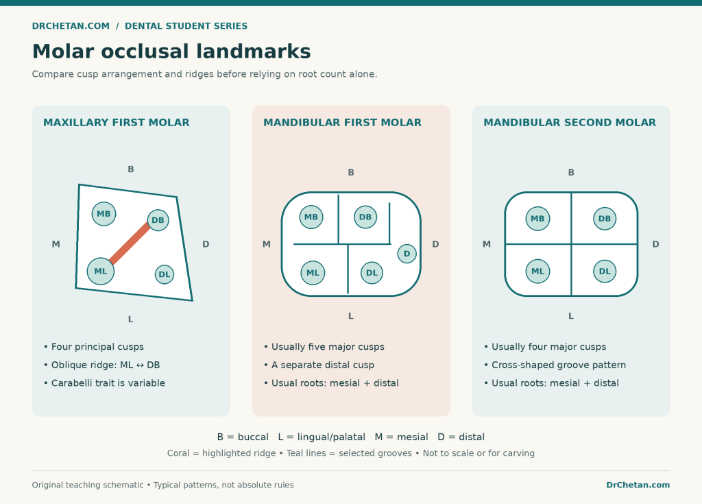
Figure 3. Occlusal landmark schematics, not carving templates or scale drawings. B = buccal, L = lingual/palatal, M = mesial, D = distal. Read each panel’s labels rather than inferring patient right or left.
Maxillary versus mandibular molars
| Feature | Typical maxillary first/second molar | Typical mandibular first/second molar |
|---|---|---|
| Root arrangement | Three: two buccal and one palatal | Two: mesial and distal |
| Crown emphasis | Greater buccolingual dimension is common | Greater mesiodistal dimension is common |
| Distinctive ridge clue | Oblique ridge between ML and DB cusps | No equivalent characteristic maxillary oblique ridge |
| First molar | Four major cusps; variable Carabelli trait | Usually five major cusps |
| Second molar | Distolingual cusp reduction can produce a heart-shaped form | Usually four cusps with a cross-shaped groove pattern |
How to assess right and left without reversing the tooth
First determine mesial and distal; then determine the patient’s side. Calling a contour “right” before orienting the tooth can produce a confident but reversed answer.
For a facing-patient labial or buccal view, place the tooth as it belongs in its arch: maxillary roots point upwards and crowns downwards; mandibular roots point downwards and crowns upwards. Right and left still belong to the patient, not the observer.
Useful side clues include:
- A sharper mesioincisal versus rounder distoincisal corner in a typical maxillary central incisor.
- The usually shorter mesial cusp ridge in an unworn permanent canine.
- The mesial crown–root depression and mesial marginal groove of a maxillary first premolar.
- The mesiolingual groove of a mandibular first premolar.
- The distal cusp of a typical mandibular first molar, once the buccal and lingual surfaces have been established.
General tendencies—such as a more cervical distal contact or greater mesial CEJ curvature—can help, but their usefulness varies by tooth. Root curvature is an additional observation, not a universal side-finding rule.
A worked side example
Suppose a typical maxillary central incisor is held with its labial surface towards you, root upwards, and incisal edge downwards. Its sharper mesioincisal corner is on your right and its rounder distal corner is on your left.
The mesial surface therefore faces towards the midline on your right. In a facing-patient view, that tooth belongs on the patient’s upper right: the permanent maxillary right central incisor, FDI 11. Changing the view or turning the tooth upside down changes what appears on your right or left, which is why orientation must come first.
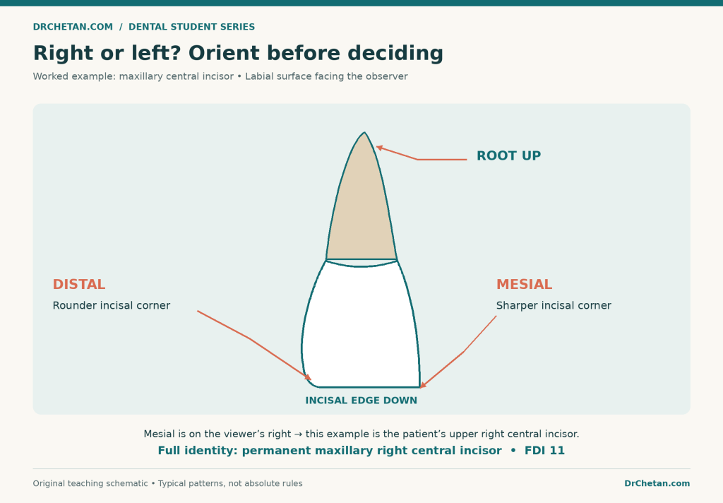
Figure 4. A deliberately simplified maxillary central-incisor example. The side is established only after arch, surface, root direction, and mesial–distal landmarks are known.
A rapid reference to the 16 permanent tooth types
Each type has right and left counterparts. The codes below are FDI identifiers for those counterparts, not a separate way to decide morphology.
| Tooth type | FDI right / left | Identification features to combine |
|---|---|---|
| Maxillary central incisor | 11 / 21 | Broad incisor crown; sharper mesial and rounder distal incisal corner |
| Maxillary lateral incisor | 12 / 22 | Smaller rounded crown; more marked lingual anatomy; variable form |
| Maxillary canine | 13 / 23 | Bulky crown; prominent labial and lingual relief; long root |
| Maxillary first premolar | 14 / 24 | Mesial marginal groove and crown–root depression; unequal cusp heights |
| Maxillary second premolar | 15 / 25 | Rounded outline; more similar cusp heights; supplementary grooves |
| Maxillary first molar | 16 / 26 | Four major cusps; oblique ridge; usual three-root arrangement |
| Maxillary second molar | 17 / 27 | Reduced distal region, especially DL cusp; possible heart-shaped form |
| Maxillary third molar | 18 / 28 | Variable reduced crown and root form; compare with other maxillary molars |
| Mandibular central incisor | 41 / 31 | Small narrow crown; relative symmetry; subdued lingual anatomy |
| Mandibular lateral incisor | 42 / 32 | Slightly larger and less symmetrical; distolingual incisal twist |
| Mandibular canine | 43 / 33 | Slender crown; smoother lingual anatomy; straighter mesial crown–root outline |
| Mandibular first premolar | 44 / 34 | Dominant buccal cusp; tiny lingual cusp; mesiolingual groove |
| Mandibular second premolar | 45 / 35 | Developed lingual part; two- or three-cusp form; H/U or Y pattern |
| Mandibular first molar | 46 / 36 | Usually five cusps; mesial and distal roots; distal cusp |
| Mandibular second molar | 47 / 37 | Usually four cusps and cross-shaped grooves; mesial and distal roots |
| Mandibular third molar | 48 / 38 | Variable crown and roots; compare with other mandibular molars |
Common confusions and how to resolve them
Canine versus mandibular first premolar: inspect the occlusal surface. A small lingual cusp, transverse ridge, and premolar table can be missed when you rely on the buccal silhouette.
Maxillary first versus second premolar: examine the mesial marginal ridge and crown–root depression, then compare cusp heights and the occlusal outline. Root count is insufficient by itself.
Mandibular central versus lateral incisor: compare symmetry and inspect the incisal view for a distolingual twist. Size is supportive, especially when comparing matched teeth, but not conclusive across unrelated specimens.
Maxillary second versus third molar: both may be heart-shaped and have closely placed or fused roots. State the whole feature combination and recognise the uncertainty of a highly variable isolated specimen.
Mandibular first versus second molar: compare the fifth distal cusp, buccal groove arrangement, and overall occlusal pattern. Do not count a worn ridge or restoration as an extra developmental cusp.
A four-cusp upper versus lower molar: look for the upper molar’s oblique ridge and root arrangement. “Four cusps” can fit more than one tooth.
How to present an answer in a practical or viva
Use an observation ? interpretation ? identification sequence. For example:
“The specimen has a broad posterior crown, five major cusps including a distal cusp, and two roots arranged mesially and distally. These features favour a permanent mandibular first molar. I would establish the buccal surface and distal cusp to assess the side.”
If you have not established the side, stop there rather than inventing one. If you have established it, give the full name and an explicitly labelled notation, such as FDI 46. Keep root and canal counts separate, and acknowledge damage or wear that limits the conclusion.
A useful practice routine
Study a known tooth from all five relevant views, then compare it directly with its nearest look-alike. Write three positive features and one feature that would make you reconsider the answer. Finally, cover the label and repeat the identification aloud.
For models, compare the same material and scale where possible. For extracted specimens, follow your institution’s handling and infection-control instructions. Do not interpret a difference in colour, stain, or tray arrangement as a diagnostic anatomical feature.
Make one pass identifying only tooth classes, a second distinguishing arches, and a third identifying individual members. Add side determination after those decisions become reliable. This isolates the source of an error instead of making every wrong answer feel like a failure to remember everything.
Practice questions
These are original revision scenarios. They ask for the most likely identification from a feature combination, not a promise that a short description can replace examination of a real specimen.
| No. | Question |
|---|---|
| 1 | A broad incisor has a sharper mesioincisal and rounder distoincisal corner. Which maxillary incisor is favoured? |
| 2 | Which permanent incisor is especially associated with a relatively small, bilaterally symmetrical crown? |
| 3 | Which incisor commonly shows a distolingual incisal twist? |
| 4 | What additional view helps distinguish a canine-like mandibular first premolar from a canine? |
| 5 | A premolar has a mesial marginal groove, mesial crown–root depression, and taller buccal cusp. Which tooth is favoured? |
| 6 | Does a single root exclude a maxillary first premolar? |
| 7 | Which premolar typically combines a dominant buccal cusp, tiny lingual cusp, and mesiolingual groove? |
| 8 | Which premolar may have three cusps and a Y-shaped groove pattern? |
| 9 | What groove patterns are commonly described for the two-cusp mandibular second premolar? |
| 10 | Which two cusps are connected by the characteristic maxillary molar oblique ridge? |
| 11 | A typical lower molar has five major cusps and mesial/distal roots. Which tooth is favoured? |
| 12 | A typical lower molar has four major cusps and a cross-shaped groove pattern. Which tooth is favoured? |
| 13 | Does absence of a conspicuous Carabelli cusp exclude a maxillary first molar? |
| 14 | Is every heart-shaped maxillary molar a third molar? |
| 15 | Why should root curvature not be your only side-identification clue? |
| 16 | In a facing-patient labial view, how do you orient a maxillary tooth’s root and crown? |
| 17 | A tooth has three roots. Does that automatically mean three root canals? |
| 18 | What is the FDI code for the permanent mandibular right first premolar? |
Answers with explanations
- Maxillary central incisor. Its broader crown and typical mesial–distal corner difference are a useful combination.
- Mandibular central incisor. The symmetry helps identify the type but can make side determination difficult.
- Mandibular lateral incisor. Inspect it from the incisal view and seek supporting asymmetry.
- The occlusal view. Look for a small lingual cusp, transverse ridge, and posterior occlusal architecture.
- Maxillary first premolar. The mesial groove and crown–root depression are particularly helpful together.
- No. Root configurations vary; reassess the crown and mesial landmarks.
- Mandibular first premolar. This combination is more useful than its canine-like buccal appearance alone.
- Mandibular second premolar. The three-cusp form has a buccal and two lingual cusps.
- H or U patterns. A mandibular second premolar does not have to be three-cusped.
- Mesiolingual and distobuccal. Check the labelled cusp positions before identifying the oblique ridge.
- Mandibular first molar. The five-cusp form includes a distal cusp; variations and wear require care.
- Mandibular second molar. Combine the regular four-cusp pattern with the other crown and root features.
- No. The Carabelli trait varies in expression and is not one of the four principal cusps.
- No. A maxillary second molar can have a reduced distolingual cusp and heart-shaped outline.
- Curvature is variable and may be altered or absent. Crown landmarks and correct orientation provide additional evidence.
- Root upwards, crown/incisal edge downwards. Establish the facial surface and mesial–distal landmarks before assigning the patient’s side.
- No. External root number and internal canal number are different anatomical observations.
- FDI 44. Quadrant 4 is permanent lower right; position 4 is the first premolar.
Final revision checklist
Ask: What class? Which arch? Which member? Which side? Which notation? For every answer, give at least a few mutually consistent observations. Check the closest alternative, then state the limits created by wear or variation.
The aim is to explain why a specimen fits a tooth type. When you can describe the shape, identify the landmarks, and show how the evidence fits together, identification becomes a reasoned process rather than a guess based on one memorised feature.

Leave a Reply