
An outdoor worker has long-standing scaling and a blurred border on the lower lip. At a later visit, one area becomes firm and repeatedly crusts. The diffuse changes can fit actinic cheilitis, while the new focus may require assessment for invasive squamous cell carcinoma. Dental students need to describe the surface of origin, record the pattern and recognise which features call for prompt referral.
Establish where on the lip the change began
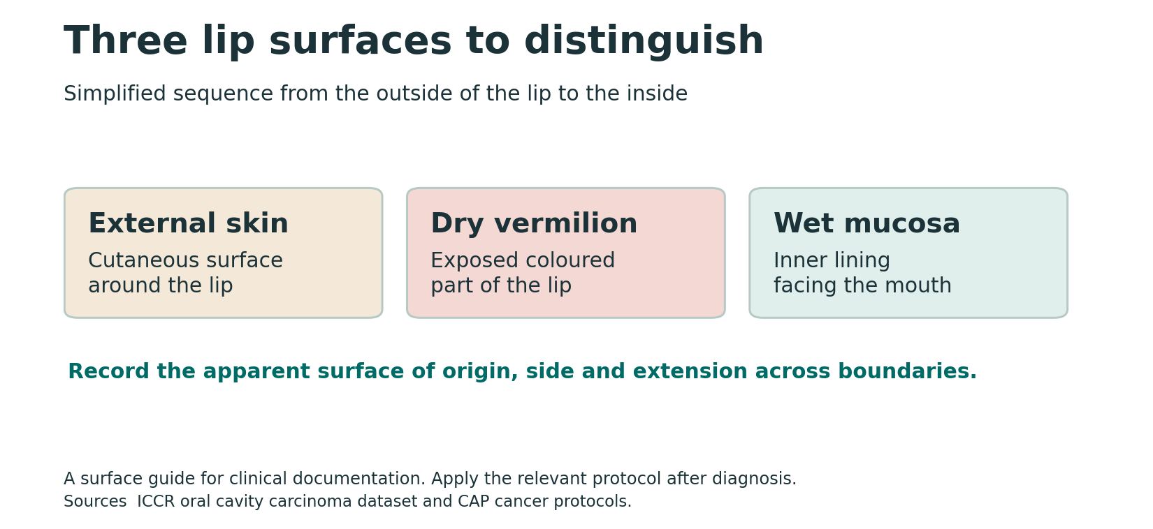
The lip has an external cutaneous surface, a dry exposed vermilion and a wet inner mucosal surface. Actinic cheilitis is associated with chronic ultraviolet exposure and commonly involves the lower vermilion. Describe where the lesion appears to originate and whether it extends onto adjacent skin or wet mucosa. If the boundary is obscured, record that uncertainty and how much of the lip is involved.
This matters if carcinoma is diagnosed. The oral cavity reporting dataset includes mucosal lip. Squamous carcinoma arising on the dry vermilion is generally handled within the cutaneous head and neck framework under the applicable protocol. A lesion that crosses the border can require clinicopathological correlation before a final site is assigned. Record the surface rather than applying a staging framework from the word “lip” alone.
Recognise diffuse change and examine for a focal lesion
| Finding | What to document | Clinical relevance |
|---|---|---|
| Dryness, scaling or surface roughness | Extent, duration and whether diffuse or localised | Can occur with chronic sun damage and with other lip disorders. |
| Blurred vermilion border or colour change | Upper or lower lip, side and relationship to adjacent skin and wet mucosa | Helps describe distribution and the likely surface of origin. |
| Focal persistent crust or ulcer | Size, exact location, duration and bleeding | A persistent focal change may need tissue assessment. |
| Palpable induration, thickening or nodule | Location, firmness, fixation and interval growth | Raises concern for invasive disease and warrants prompt assessment when suspicious. |
Ask about years of outdoor work or recreation, regular sun protection and prior lip lesions or procedures. Examine the full vermilion and adjacent skin and mucosa. Palpate a focal change where appropriate and include the neck assessment when findings warrant it. Record whether there is a single dominant focus or widespread change. Photograph with consent and scale if helpful, while giving the location in words.
Dry, cracked or inflamed lips can have several causes. Irritant or allergic cheilitis, trauma, infection and inflammatory disorders can overlap in appearance. An angular fissure has a different distribution from a chronic diffuse change along the lower vermilion. A typical exposure history can support the assessment, but the time course and focal signs should determine whether a lesion needs further investigation.
Decide what needs tissue assessment

Actinic cheilitis is included among oral potentially malignant disorders. The clinical appearance does not reliably establish the degree of epithelial change or exclude a focus of invasion. A persistent focal ulcer, progressive thickening, recurrent bleeding, induration or a growing nodule deserves prompt specialist assessment or biopsy by an appropriately trained clinician. When diffuse change contains a more suspicious focus, identify that area explicitly on the referral.
A specialist can decide whether to sample a focal area or more than one site and how to obtain adequate tissue. When planning treatment, the German S3 guideline recommends biopsy before treatment to confirm the diagnosis and exclude invasive SCC. Its recommendations come from a particular guideline context. The relevant principle at chairside is to arrange assessment before proceeding with treatment that might remove or obscure the suspicious tissue without providing a diagnosis.
If a biopsy shows chronic sun-related change or dysplasia, compare the sampled site with the visible and palpable findings. If the most suspicious region was not sampled or the clinical findings progress, discuss further assessment. Where invasion is confirmed, document the precise surface of origin so the pathology and oncology teams can apply the appropriate staging and treatment framework.
Write a referral with an identifiable target
Give the receiving clinician a location that can be checked at the next visit, such as left lower dry vermilion near the commissure, extending toward the wet mucosa. Document the dimensions, firmness and whether the change is within more diffuse scaling or at a separate site. If the surface is crusted, explain whether there is a persistent palpable change beneath it. Include prior pathology and the reason a focal assessment is needed.
Ask about prior lip treatment, occupational sun exposure and relevant immunosuppression when taking the history. These factors help the specialist assess risk and select management. The existing clinical findings still determine whether a focal change needs prompt investigation. A referral that names only actinic cheilitis can miss the reason for concern, especially when the patient already knows that diagnosis.
Interpret transformation estimates cautiously
A frequently cited systematic review reported a transformation proportion of 3.07%, based on the single study that met its inclusion criteria. That estimate depends on the cohort, case definition and follow-up. Other reviews have used different eligibility and outcome definitions. It is more useful to tell a patient that actinic cheilitis can precede SCC, that a suspicious focus may already need investigation and that the plan depends on what the clinician observes and the biopsy establishes.
Baseline carcinoma and cancer developing later are separate outcomes. A lip with chronic scaling can contain a new invasive focus at the first specialist assessment. That is different from following a lesion that had no invasion at baseline and observing a future cancer. When comparing publications, look for whether they excluded initial SCC, how they confirmed diagnoses and how long they followed patients.
Discuss prevention, treatment and follow-up as separate steps
Reducing ultraviolet exposure is relevant to prevention and continued care. Discuss practical protection for the lips and face, such as shade, a broad-brimmed hat and appropriate lip sun protection. A prevention discussion can happen while a focal lesion is being investigated. It does not replace biopsy or referral for an area that is suspicious.
Specialists select management according to the clinical extent, histology, functional and cosmetic considerations, and patient preferences. Options may include procedures to remove or treat involved vermilion. The best choice for a small focal area may differ from extensive field change. A follow-up plan should identify who will reassess the treated area, check pathology if obtained and evaluate new focal changes.
If a biopsy has already been performed, ask which part of the lip was sampled and whether it included the focal finding. When a procedure treats a broad area, establish how the specialist will document healing and when the patient should report a new firm or ulcerated focus. A photograph taken with consent can be useful when the treated border is difficult to describe in words alone.
Apply the distinction at chairside
Diffuse scaling without a dominant focus. Record the exposure history, distribution and relevant findings. Discuss practical protection and arrange appropriate assessment or follow-up according to the clinical impression. A baseline description helps identify whether a later focus is new.
New induration within chronic vermilion change. Describe the side, dimensions, palpable firmness and relationship to the dry and wet lip. Arrange prompt assessment of the focal change. Give the specialist the previous record if available, and check that the patient reached the intended service.

Leave a Reply