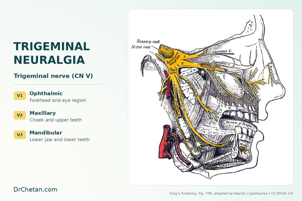
Trigeminal neuralgia can feel as if it starts in a tooth even when the teeth cannot account for the attacks. A person may report a sudden electric shock across the cheek or jaw when brushing, washing the face or touching the lip. The dental assessment has two jobs. Check for disease that explains the pain, and recognise a pattern that calls for neurological or specialist orofacial assessment before irreversible treatment.
Recognise the pattern over time
The International Classification of Headache Disorders describes recurrent, usually unilateral attacks in one or more divisions of the trigeminal nerve. Individual attacks typically last from a fraction of a second to two minutes. The pain is severe, with a sharp, shooting or electric shock-like quality. An innocuous stimulus within the affected territory, such as a light touch, can trigger attacks. A person can have many attacks in a cluster. Some also have continuous or near-continuous pain in the same territory between them.
Ask the patient to distinguish the length of one shock from the length of the whole episode. Ask where the shock begins and travels, whether it crosses the midline, which activities trigger it, and what they feel between attacks. Upper jaw pain can fall within the maxillary division, V2. Lower jaw or lip pain can fall within the mandibular division, V3. A map of the pain and triggers is useful for referral. Repeatedly provoking a severe attack in clinic adds little when the history is already clear.
Check for a cause in the mouth
| Pattern | What to compare |
|---|---|
| Brief shocks triggered by light touch | Record distribution, attack duration and intervals. Check whether dental examination offers an explanation. |
| Tooth pain with thermal or biting symptoms | Compare pulp sensibility, percussion, bite testing, periodontal findings and appropriate imaging with the history. |
| Persistent burning with altered sensation after a procedure | Map any sensory change and consider painful post-traumatic trigeminal neuropathy in the differential. |
| Ache with jaw movement or muscle palpation | Examine masticatory muscles and the temporomandibular joints for reproduction of the familiar pain. |
Tests need to agree with the patient’s complaint. A heavily restored tooth or minor radiographic finding may be incidental. Dental disease and neuralgia can also coexist. If one tooth has a clear problem, explain which symptoms it is likely to account for and which still require assessment. Deferring an extraction or root canal is appropriate when there is no diagnosis that supports it.
Classification and imaging
Classical trigeminal neuralgia is associated with neurovascular compression producing a morphological change in the nerve, such as displacement or atrophy. Simple vessel–nerve contact can be an incidental finding. Secondary trigeminal neuralgia is attributable to another disorder, such as multiple sclerosis or a lesion affecting the nerve. Idiopathic trigeminal neuralgia is used when adequate investigation fails to identify evidence meeting the criteria for those categories. MRI forms part of the specialist work-up, including assessment for a secondary cause. Dental radiographs help assess the teeth and jaws and answer a different question.
A sensory deficit, bilateral symptoms, persistent atypical pain or another neurological finding should raise concern and be communicated promptly. Record the onset and evolution of symptoms, oral and sensory findings, and any prior treatment. Severe attacks can interfere with drinking, eating and oral hygiene, which should also be included in the referral.
What treatment can involve
Carbamazepine or oxcarbazepine are usual first-line medicines in specialist guidance. The prescriber needs to review interactions, medical history and appropriate baseline or follow-up monitoring. If pain remains difficult to control or the medicines are poorly tolerated, specialist teams can consider procedures. Microvascular decompression is one option in selected classical cases. Other procedures include lesioning techniques and stereotactic radiosurgery, depending on the findings, preferences and individual risks.
A dental example
A patient reports half-second shocks across the right lower lip and jaw when the toothbrush touches the lip. A lower molar has a large filling. Its examination and imaging give no convincing explanation for the shocks. Record the time course, distribution and triggers and arrange an appropriate referral. Replacing the filling or extracting the tooth without a corroborating dental diagnosis could leave the original pain untreated.

Leave a Reply